. 2016 Sep 7;1(5):e00235-16.
doi: 10.1128/mSphere.00235-16. eCollection 2016 Sep-Oct.

Biochemical Characterization of Middle East Respiratory Syndrome Coronavirus Helicase

Affiliations

Biochemical Characterization of Middle East Respiratory Syndrome Coronavirus Helicase

Adeyemi O Adedeji et al. mSphere. .

Abstract

Middle East respiratory syndrome coronavirus (MERS-CoV) helicase is a superfamily 1 helicase containing seven conserved motifs. We have cloned, expressed, and purified a Strep-fused recombinant MERS-CoV nonstructural protein 13 (M-nsp13) helicase. Characterization of its biochemical properties showed that it unwound DNA and RNA similarly to severe acute respiratory syndrome CoV nsp13 (S-nsp13) helicase. We showed that M-nsp13 unwound in a 5'-to-3' direction and efficiently unwound the partially duplex RNA substrates with a long loading strand relative to those of the RNA substrates with a short or no loading strand. Moreover, the Km of ATP for M-nsp13 is inversely proportional to the length of the 5' loading strand of the partially duplex RNA substrates. Finally, we also showed that the rate of unwinding (ku) of M-nsp13 is directly proportional to the length of the 5' loading strand of the partially duplex RNA substrate. These results provide insights that enhance our understanding of the biochemical properties of M-nsp13. IMPORTANCE Coronaviruses are known to cause a wide range of diseases in humans and animals. Middle East respiratory syndrome coronavirus (MERS-CoV) is a novel coronavirus discovered in 2012 and is responsible for acute respiratory syndrome in humans in the Middle East, Europe, North Africa, and the United States of America. Helicases are motor proteins that catalyze the processive separation of double-stranded nucleic acids into two single-stranded nucleic acids by utilizing the energy derived from ATP hydrolysis. MERS-CoV helicase is one of the most important viral replication enzymes of this coronavirus. Herein, we report the first bacterial expression, enzyme purification, and biochemical characterization of MERS-CoV helicase. The knowledge obtained from this study might be used to identify an inhibitor of MERS-CoV replication, and the helicase might be used as a therapeutic target.

Keywords: ATP hydrolysis; DNA; RNA; coronavirus; enzyme kinetics; helicase.


Figures

FIG 1
FIG 1
Protein expression and purification. (A) M-nsp13 and FMDV polymerase (pol) genes were cloned in pET52b and pet-28b, respectively, followed by their expression in the E. coli BL21 bacterial expression system. M-nsp13 was then purified by Strep-Tactin affinity chromatography. The FMDV polymerase was purified using nickel affinity chromatography. Purified M-nsp13 and FMDV polymerase were readily visualized by Coomassie blue staining of SDS-PAGE gels as ~68-kDa and 55-kDa protein products, respectively, in line with their expected molecular masses. (B) Western immunoblot analysis with M-nsp13-specific rabbit antiserum. The positions of protein molecular mass markers are indicated on the left (in kilodaltons).
FIG 2
FIG 2
Sequence comparison of coronavirus helicases. The alignment was generated with the UniProt program (http://www.uniprot.org/align/) and the nsp13 sequences of MERS-CoV (human betacoronavirus strain 2c EMC/2012; GenBank accession no. JX869059.2), SARS-CoV (isolate Frankfurt 1; accession no. AY291315), mouse hepatitis virus (MHV; strain A59; accession number NC_001846), transmissible gastroenteritis virus (TGEV; strain Purdue 46; accession number AJ271965), and avian infectious bronchitis virus (IBV; strain Beaudette; accession number M95169) were derived from the replicative polyproteins of these viruses, whose sequences were obtained from the GenBank database. Conserved helicase motifs I to VI are indicated. Also indicated by an @ sign is the conserved Lys288 residue (corresponding to Lys5598 in pp1ab), which in the M-nsp13_K288A control protein was replaced with Ala. Lys288 is part of a conserved helicase motif (I) which is also called the Walker A box. Near the N terminus, the 12 conserved Cys and His residues predicted to form a binuclear zinc-binding cluster are indicated by a circled “C.” Overall, MERS helicase has 72.4% identity with SARS, 67.2% identity with MHV, 61.3% identity with TGEV, and 59.1% with IBV helicases.
FIG 3
FIG 3
Helicase activity of M-nsp13. (A) Determination of optimal enzyme concentration. The activity of purified M-nsp13 was determined in an enzyme-dependent manner using a 5′-Cy3-labeled (*) partially duplex 5′-RNA-20 (20 ss, 22 ds) RNA substrate for 30 min. The reaction products were separated on a nondenaturing 8% polyacrylamide gel and visualized using the Bio-Rad multipurpose imager. (B) Comparison of fractions of partially duplex RNA and DNA substrates unwound by M-nsp13. The activity of purified M-nsp13 was verified in a time-dependent manner using a 5′-Cy3-labeled (*) partially duplex 5′-RNA-20 (20 ss, 22 ds) RNA substrate (upper gel) and a 5′-DNA-20 (20 ss, 22 ds) DNA substrate (lower gel). The reaction products were separated on a nondenaturing 8% polyacrylamide gel and visualized using the Bio-Rad multipurpose imager.
FIG 4
FIG 4
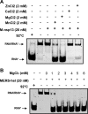
Determination of metal requirement. (A) The activity of purified M-nsp13 was performed using a 5′-Cy3-labeled (*) partially duplex 5′-RNA-20 (20 ss, 22 ds) RNA substrate in the presence of 2 mM of Zn2+, Ca2+, Mg2+, and Mn2+. The reaction products were separated on a nondenaturing 8% polyacrylamide gel and visualized using the Bio-Rad multipurpose imager. (B) Purified M-nsp13 was incubated with the 5′-Cy3-labeled (*) partially duplex 5′-RNA-20 (20 ss, 22 ds) RNA substrate in the presence of various concentrations of Mg. The reaction products were separated on a nondenaturing 8% polyacrylamide gel and visualized using the Bio-Rad multipurpose imager. hel, helicase.
FIG 5
FIG 5
pH dependence. Purified M-nsp13 was incubated with the 5′-Cy3-labeled (*) partially duplex 5′-RNA-20 (20 ss, 22 ds) RNA substrate in reaction buffers with different pHs (4.0 to 9.0). The reaction products were separated on a nondenaturing 8% polyacrylamide gel and visualized using the Bio-Rad multipurpose imager.
FIG 6
FIG 6
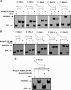
Substrate specificity, minimum overhang length, and ATP requirement for M-nsp13. (A and B). Five different substrates with 5′-overhang lengths varying from 0 to 20 nucleotides were designed to determine the minimum length of the loading strand required by M-nsp13 to efficiently unwind its substrate. The helicase activity of M-nsp13 (20 nM) was assessed on these substrates (5 nM each) at 30°C with 2 mM ATP (A) and no ATP (B). The products were separated on a nondenaturing 8% polyacrylamide gel and visualized using the Bio-Rad multipurpose imager. (C) Purified M-nsp13 or M-nsp13_K288A (20 nM) was incubated with the 5′-Cy3-labeled (*) partially duplex 5′-RNA-20 (20 ss, 22 ds) RNA substrate (5 nM) in the presence of 2 mM ATP. The reaction products were separated on a nondenaturing 8% polyacrylamide gel and visualized using the Bio-Rad multipurpose imager.
FIG 7
FIG 7
(d)NTP requirements for MERS helicase-unwinding activities. (A) Five different 5′-Cy3-labeled (*) partially duplex RNA substrates with 5′-overhang lengths varying from 0 to 20 nucleotides, i.e., 0 ss, 22 ds (○), 2 ss, 22 ds (Δ), 5 ss, 22 ds (▼), 10 ss, 22 ds (◇), and 20 ss, 22 ds (■) RNA substrates, were reacted with M-nsp13 (20 nM) in the presence or absence of increasing concentrations (0 to 5 mM) of ATP for 30 min. The reaction products were separated on a nondenaturing 8% polyacrylamide gel and visualized using the Bio-Rad multipurpose imager. The unwinding activities under different ATP concentrations were plotted as the fraction of the RNA released from the total RNA helix substrate (y axis) at each ATP concentration (x axis) using the Michaelis-Menten equation. The values for Km and Vmax are provided in Table 1. Error bars represent standard deviation (SD) values from two separate experiments. (B) The 5′-Cy3-labeled (*) partially duplex 5′-RNA-10 (10 ss, 22 ds) RNA substrate (5 nM) was reacted with M-nsp13 (20 nM) in the presence or absence of the indicated NTPs (2 mM) for 30 min. The reaction products were separated on a nondenaturing 8% polyacrylamide gel and visualized using the Bio-Rad multipurpose imager.
FIG 8
FIG 8
M-nsp13 rates of unwinding of all RNA substrates with 5′ loading strands of various lengths. (A) Five different 5′-Cy3-labeled (*) partially duplex RNA substrates with 5′-overhang lengths varying from 0 to 20 nucleotides, i.e., 0 ss, 22 ds (○), 2 ss, 22 ds (Δ), 5 ss, 22 ds (▼), 10 ss, 22 ds (◇), and 20 ss, 22 ds (■), were reacted with M-nsp13 (20 nM) in the presence of 2 mM ATP for 0 to 45 min. The reaction products were separated on a nondenaturing 8% polyacrylamide gel and visualized using the Bio-Rad multipurpose imager. The derived fractions of RNA unwound at the indicated time points were plotted against time (in minutes), and the data points were fit to a single-exponential equation (see Results) to determine the rates of unwinding (ku) of all substrates (Table 2). Error bars represent SD from two separate experiments.
FIG 9
FIG 9
M-nsp13 does not unwind partially duplex RNA substrates with a 3′ loading strand. Four different substrates with 3′-overhang lengths varying from 2 to 20 nucleotides were designed to determine whether M-nsp13 can unwind in a 3′-to-5′ direction. The helicase activity of M-nsp13 (20 nM) was assessed on these substrates (5 nM each) at 30°C for 5 min in the presence of 2 mM ATP. The products were separated on a nondenaturing 8% polyacrylamide gel and visualized using the Bio-Rad multipurpose imager.

References

    1. Adedeji AO, Singh K, Kassim A, Coleman CM, Elliott R, Weiss SR, Frieman MB, Sarafianos SG. 2014. Evaluation of SSYA10-001 as a replication inhibitor of severe acute respiratory syndrome, mouse hepatitis, and Middle East respiratory syndrome coronaviruses. Antimicrob Agents Chemother 58:4894–4898. doi:10.1128/AAC.02994-14. - DOI - PMC - PubMed
    1. Raj VS, Mou H, Smits SL, Dekkers DH, Müller MA, Dijkman R, Muth D, Demmers JA, Zaki A, Fouchier RA, Thiel V, Drosten C, Rottier PJ, Osterhaus AD, Bosch BJ, Haagmans BL. 2013. Dipeptidyl peptidase 4 is a functional receptor for the emerging human coronavirus-EMC. Nature 495:251–254. doi:10.1038/nature12005. - DOI - PMC - PubMed
    1. Raj VS, Osterhaus AD, Fouchier RA, Haagmans BL. 2014. MERS: emergence of a novel human coronavirus. Curr Opin Virol 5:58–62. doi:10.1016/j.coviro.2014.01.010. - DOI - PMC - PubMed
    1. Hocke AC, Becher A, Knepper J, Peter A, Holland G, Tönnies M, Bauer TT, Schneider P, Neudecker J, Muth D, Wendtner CM, Rückert JC, Drosten C, Gruber AD, Laue M, Suttorp N, Hippenstiel S, Wolff T. 2013. Emerging human Middle East respiratory syndrome coronavirus causes widespread infection and alveolar damage in human lungs. Am J Respir Crit Care Med 188:882–886. doi:10.1164/rccm.201305-0954LE. - DOI - PubMed
    1. Zhang N, Jiang S, Du L. 2014. Current advancements and potential strategies in the development of MERS-CoV vaccines. Expert Rev Vaccines 13:761–774. doi:10.1586/14760584.2014.912134. - DOI - PMC - PubMed

LinkOut - more resources