Observational Study
. 2016 Sep;95(37):e4902.
doi: 10.1097/MD.0000000000004902.

Different surgical strategies of patients with intravenous leiomyomatosis

Affiliations
Observational Study

Different surgical strategies of patients with intravenous leiomyomatosis

Guotao Ma et al. Medicine (Baltimore). 2016 Sep.

Abstract

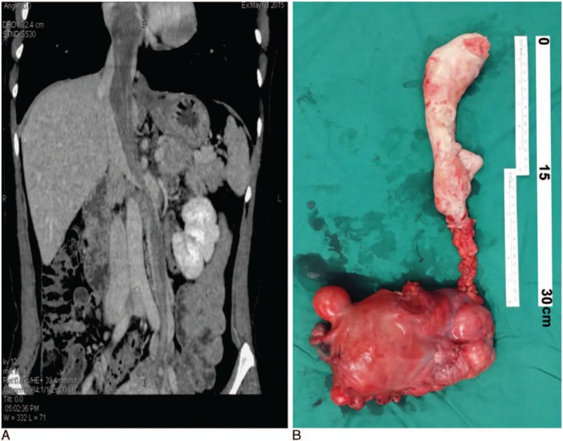
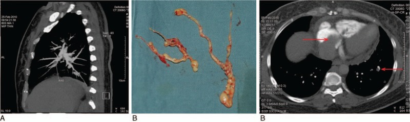
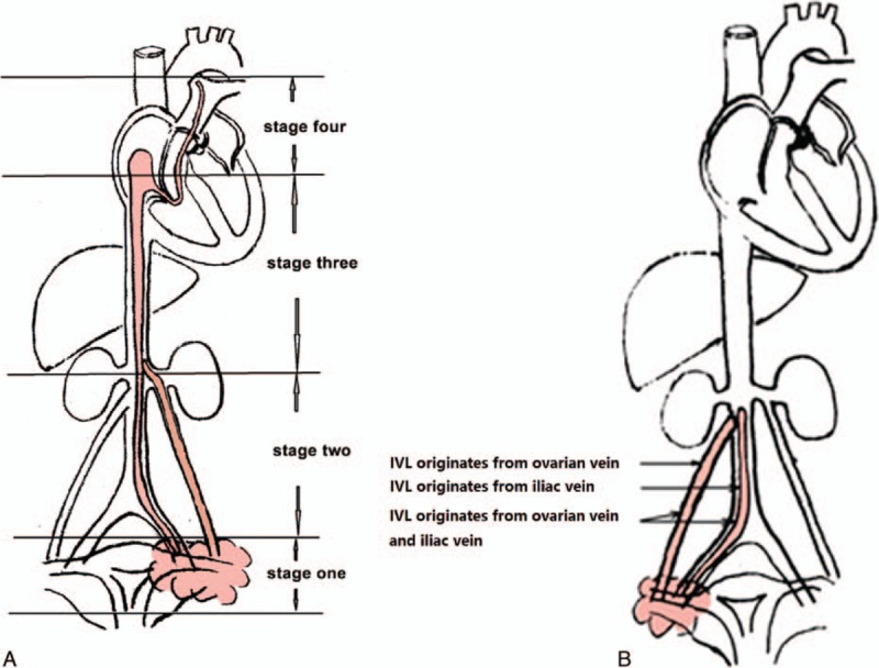
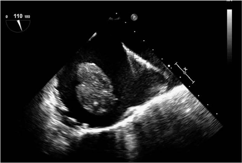
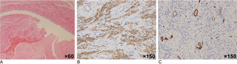
Intravenous leiomyomatosis (IVL) is a rare benign tumor. The study aimed to assess outcomes of patients treated surgically for IVL.Between November 2002 and January 2015, 76 patients were treated for IVL. The stage of IVL was evaluated preoperatively by echocardiography and enhanced computerized tomography (CT) scan, and graded into 4 stages according to intravascular tumor progression. We recorded age, lower limb edema before surgery, surgical parameters, and hospitalization expenses. Patients were followed up every 6 months and tumor recurrence was assessed by CT and ultrasound. Patients were followed up for a mean of 4.5 ± 2.5 years (range 1-13 years) and there was no operative, hospital, or long-term mortality or were lost to follow-up.The rate of lower extremity edema, amount of blood loss, postoperative transfusion, length of intensive care unit (ICU) stay, postoperative hospitalization, and hospitalization expenses differed significantly between patients at different presurgery stages. Tumors recurred in 4 of 7 patients with stage I IVL that opted for surgery that preserved the ovaries and uterus. No recurrence was observed in patients graded stage II or more, in all of which the uterus and ovaries were removed. Recurrence was observed in only 4 of 76 cases of IVL, all of whom opted for surgery that spared the ovaries and uterus.Different surgical strategies should be decided based on the staging to completely remove the tumor and ensure the safety of patients. Removal of both ovaries is necessary for inhibiting tumor growth and avoiding recurrence.


Conflict of interest statement

The authors have no conflicts of interest to disclose.

Figures

Figure 1
Figure 1
An example of stage III patient. A 44-year-old patient with stage III, IVL complained of chest tightness and shortness of breath. An enhanced CT scan was performed a week before surgery (A), and the patient underwent cardiopulmonary bypass-assisted surgery, with deep hypothermia circulation arrest. The right atrium and inferior vena cava were opened and the uterus, ovary, and tumor embolus was removed using an abdominal thoracic incision (B).
Figure 2
Figure 2
An example of stage IV patient. A 48-year-old patient, with stage IV IVL complained of chest pain and palpitations. An enhanced CT scan of the pulmonary artery performed a week before surgery (A) revealed a tumor embolus (arrow) in the right pulmonary branch. The patient underwent cardiopulmonary bypass-assisted surgery with deep hypothermia circulation arrest. The right atrium, pulmonary artery, and inferior vena cava were opened and the tumor embolus in the pulmonary artery was removed using a horizontal position, abdominal thoracic incision (B). An enhanced CT scan of the chest revealed a pulmonary metastasis. The tumor embolus was located within the right atrium, right ventricle, and a pulmonary metastasis was identified (arrow) (C).
Figure 3
Figure 3
Stage and origin of intravenous leiomyomatosis. Intravenous leiomyomatosis was categorized into 4 stages reflecting presurgery tumor progression (A), and was found to originate from different tissues (B).
Figure 4
Figure 4
Transesophageal echocardiography (TEE) revealed a strip-type mass in the right atrium of a stage III patient. A 55-year-old patient with stage III IVL complained of palpitations and shortness of breath. The patient underwent cardiopulmonary bypass-assisted surgery with deep hypothermia circulation arrest. Intraoperative TEE revealed a strip-type mass, extending from the inferior vena cava to right atrium. The head end was enlarged to 66 mm × 25 mm, and it entered into right ventricular while ventricular diastole. The right atrium and inferior vena cava were opened and the tumor was removed via an abdominal thoracic incision.
Figure 5
Figure 5
Tumor embolus specimens. HE staining of tumor embolus specimens indicated bland spindle cells (A, ×60), and immunohistochemical staining indicated Desmin (FB) and SMA (+) (B, ×150). The Ki-67 index was 2%, and no CD10, CD31, or CD34+ cells were detected (C, ×150).

References

    1. Birch-Hirschfeld FV. Lehrbuch der pathologischen anatomie. 1896; Leipzig, Germany: FCW Vogel, 226–258.
    1. Dürck H. Ueber ein kontinvierlich durch die learned Holvene in das Herz vorwachsendes Fibromyom des Uterus. München Med Wochenschr 1907; 54:1154.
    1. Marshall JF, Morris DS. Intravenous leiomyomatosis of the uterus and pelvis: case report. Ann Surg 1959; 149:126–134. - PMC - PubMed
    1. Choi IJ, Han MS, Cha MS. A huge case of intravenous leiomyomatosis with intracardiac extension. J Women Med 2010; 3:29–31.
    1. Li B, Chen X, Chu YD, et al. Intracardiac leiomyomatosis: a comprehensive analysis of 194 cases. Interact Cardiovasc Thorac Surg 2013; 17:132–138. - PMC - PubMed

Publication types