Case Reports
. 2017 Jan;41(1):12-24.
doi: 10.1097/PAS.0000000000000721.

Novel High-grade Endometrial Stromal Sarcoma: A Morphologic Mimicker of Myxoid Leiomyosarcoma

Affiliations
Case Reports

Novel High-grade Endometrial Stromal Sarcoma: A Morphologic Mimicker of Myxoid Leiomyosarcoma

Lien N Hoang et al. Am J Surg Pathol. 2017 Jan.

Abstract

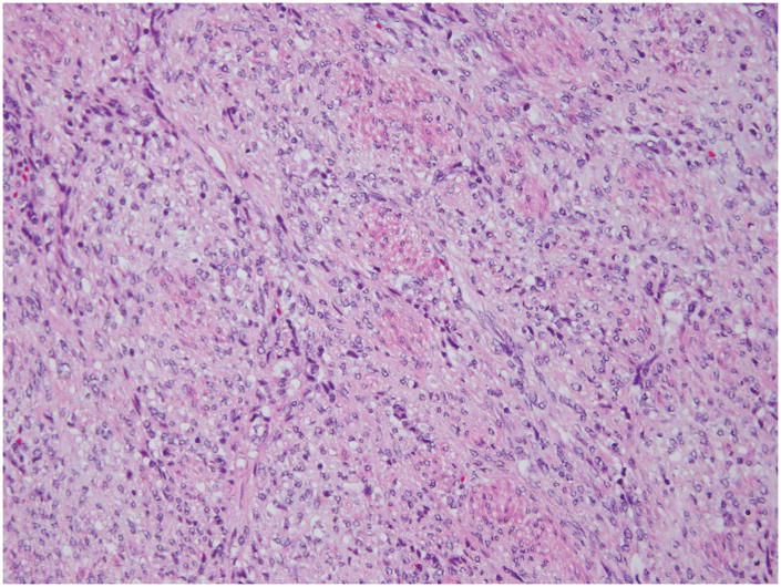
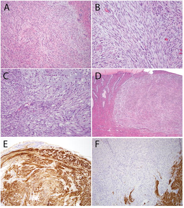
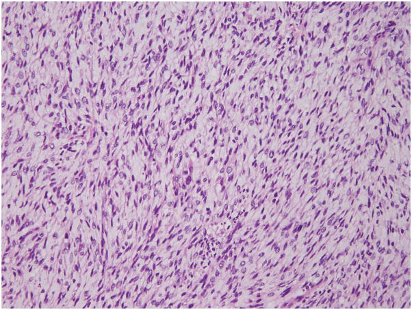
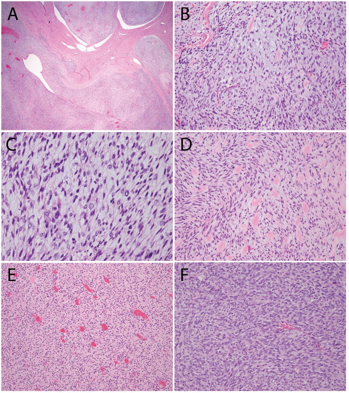
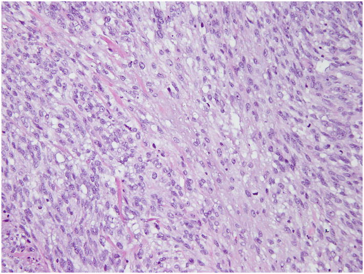
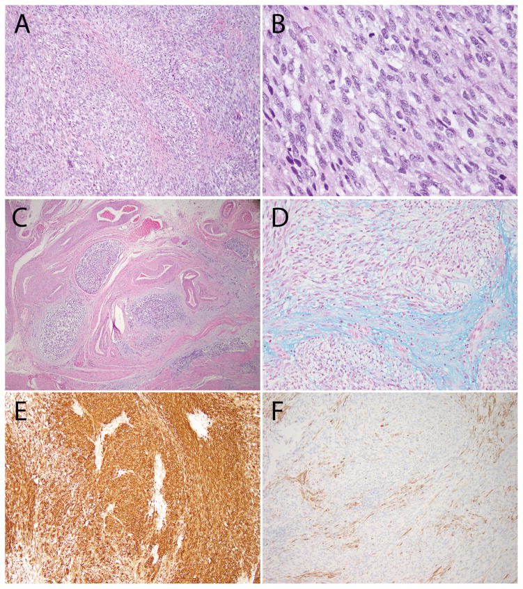
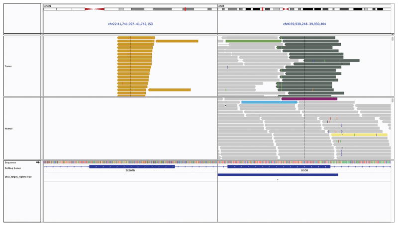
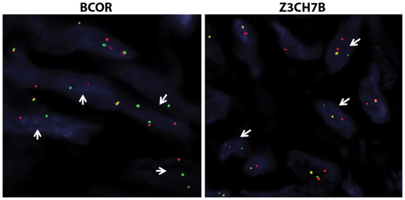
Endometrial stromal sarcomas (ESS) are often underpinned by recurrent chromosomal translocations resulting in the fusion of genes involved in epigenetic regulation. To date, only YWHAE-NUTM2 rearrangements are associated with distinctive high-grade morphology and aggressive clinical behavior. We identified 3 ESS morphologically mimicking myxoid leiomyosarcoma of the uterus and sought to describe their unique histopathologic features and identify genetic alterations using next-generation sequencing. All cases displayed predominantly spindled cells associated with abundant myxoid stroma and brisk mitotic activity. Tumors involved the endometrium and demonstrated tongue-like myometrial infiltration. All 3 were associated with an aggressive clinical course, including multisite bony metastases in 1 patient, progressive peritoneal disease after chemotherapy in another, and metastases to the lung and skin in the last patient. All 3 ESS were found to harbor ZC3H7B-BCOR gene fusions by targeted sequencing and fluorescence in situ hybridization. On the basis of the review of these cases, we find that ESS with ZC3H7B-BCOR fusion constitutes a novel type of high-grade ESS and shares significant morphologic overlap with myxoid leiomyosarcoma.


Conflict of interest statement

The authors have no conflicts of interest to disclose.

Figures

Figure 1
Figure 1. Histopathology of t(X;22) ZC3H7B-BCOR ESS, case 1
The tumor was spindled with epithelioid areas, showed focal fascicular architecture and extensive myxoid change. The tumor cell nuclei were enlarged, with irregular nuclear contours and displayed clear to hyperchromatic chromatin.
Figure 2
Figure 2. Additional histologic features of ESS with ZC3H7B-BCOR translocation, case 1
(A) Predominantly spindled neoplasm with myxoid features and a pushing border; (B) Focal fascicular architecture and thin-walled blood vessels; (C) Areas with more prominent myxoid stroma; (D) Involvement of the uterine cervix; (E) CD10 was strongly positive; (F) Desmin was negative (positive staining seen in background myometrium).
Figure 3
Figure 3. Histopathology of t(X;22) ZC3H7B-BCOR ESS, case 2
The tumor was composed of spindled and epithelioid cells with extensive myxoid changes. The tumor cell nuclei were monotonous, and the chromatin varied from pale/clear to hyperchromatic.
Figure 4
Figure 4. Additional histologic features of ESS with ZC3H7B-BCOR translocation, case 2
(A) Tongue-like myoinvasion and worm-like plugs of the uterine vessels; (B) Spindled cells embedded in myxoid stroma; (C) Cytologic atypia with enlarged nuclei, irregular nuclear contours and hyperchromatic to pale/clear chromatin; (D) Collagen plaques; (E) Thin-walled blood vessels; (F) More cellular areas were present with less myxoid stroma.
Figure 5
Figure 5. Histopathology of t(X;22) ZC3H7B-BCOR ESS, case 3
The tumor was composed of spindled cells in a background diffuse myxoid stroma. The tumor cell nuclei were enlarged, had irregular nuclear contours, coarse chromatin and some had conspicuous nucleoli. Mitoses were up to 10/10 high power fields.
Figure 6
Figure 6. Additional histologic features of ESS with ZC3H7B-BCOR translocation, case 3
(A–B) The tumor was composed of spindled cells in a myxoid stroma; (C) Worm-like plugs of tumor were seen in blood vessels; (D) Alcian blue pH 2.5 was positive; (E) CD10 was strongly and diffusely positive; (F) Caldesmon was negative (positive staining seen in background myometrium).
Figure 7
Figure 7. Pictorial representation of the ZC3H7B-BCOR translocation on Integrated Genome Viewer
Each bar is a single sequenced read with the top panel displaying data from tumor sample and bottom panel displaying its matched normal sample. The reads are sorted to highlight read pairs with discordant read mapping on the top for each sample. As evident, one set of reads maps to ZC3H7B on chromosome 22 while the their corresponding paired end reads map to BCOR on the X chromosome, supporting the presence of a somatic fusion involving both genes.
Figure 8
Figure 8. Fluorescence in-situ hybridization confirmation of BCOR and ZC3H7B rearrangements in ESS case 1
Break-apart assays for BCOR and ZC3H7B both show split signals (arrows, red probe centromeric, green probe telomeric) confirming rearrangements in both genes.

References

    1. Kosary CL. Cancer of the Corpus Uteri. In: Ries LAG, Young JL, Keel GE, et al., editors. SEER Survival Monograph: Cancer Survival Among Adults: US SEER Program, 1988–2001, Patient and Tumor Characteristics. Bethesda, MD: National Cancer Institute; 2007. pp. 123–132. SEER Program, NIH Pub. No. 07-6215.
    1. Norris HJ, Taylor HB. Mesenchymal tumors of the uterus: A clinical and pathological study of 53 endometrial stromal tumors. Cancer. 1966;19:755–766. - PubMed
    1. Lee CH, Nucci MR. Endometrial stromal sarcoma--the new genetic paradigm. Histopathology. 2015;67:1–19. - PubMed
    1. Ali RH, Rouzbahman M. Endometrial stromal tumours revisited: an update based on the 2014 WHO classification. J Clin Pathol. 2015;68:325–332. - PubMed
    1. Conklin CMJ, Longacre TA. Endometrial stromal tumors: the new WHO classification. Adv Anat Pathol. 2014;21:383–393. - PubMed

Publication types

MeSH terms