. 2017 Jan;56(1):1-10.
doi: 10.1165/rcmb.2016-0181MA.

Transplantation of Airway Epithelial Stem/Progenitor Cells: A Future for Cell-Based Therapy

Affiliations

Transplantation of Airway Epithelial Stem/Progenitor Cells: A Future for Cell-Based Therapy

Moumita Ghosh et al. Am J Respir Cell Mol Biol. 2017 Jan.

Abstract

Cell therapy has the potential to cure disease through replacement of malfunctioning cells. Although the tissue stem cell (TSC) is thought to be the optimal therapeutic cell, transplantation of TSC/progenitor cell mixtures has saved lives. We previously purified the mouse tracheobronchial epithelial TSCs and reported that in vitro amplification generated numerous TSCs. However, these cultures also contained TSC-derived progenitor cells and TSC repurification by flow cytometry compromised TSC self-renewal. These limitations prompted us to determine if a TSC/progenitor cell mixture would repopulate the injured airway epithelium. We developed a cell transplantation protocol and demonstrate that transplanted mouse and human tracheobronchial epithelial TSC/progenitor cell mixtures are 20-25% of airway epithelial cells, actively contribute to epithelial repair, and persist for at least 43 days. At 2 weeks after transplantation, TSCs/progenitor cells differentiated into the three major epithelial cell types: basal, secretory, and ciliated. We conclude that cell therapy that uses adult tracheobronchial TSCs/progenitor cells is an effective therapeutic option.

Keywords: airway epithelium; cell therapy; progenitor cell; tissue stem cell.


Figures
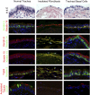
Figure 1.
Figure 1.
Mouse tissue stem cell (TSC)/progenitor cell differentiation in rat tracheal xenografts. Rat tracheal scaffolds were seeded with 1 × 106 irradiated NIH3T3 fibroblasts or viable mouse TSCs/progenitor cells and implanted in a dorsal–lateral subcutaneous pocket of nonobese diabetic (NOD)/severe combined immunodeficiency (SCID)/TSC-γ knockout mice. Xenografts and the host trachea were recovered on Day 14. Histological sections were stained as indicated. All images are ×200 magnification. Data are representative of four experiments. CCSP, Clara cell secretory protein; H&E, hematoxylin and eosin; Trp63, tumor protein 63.
Figure 2.
Figure 2.
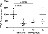
Naphthalene (NA) treatment depletes TSCs. Female FVB/n mice were treated with corn oil or 300 mg/kg NA and recovered for 6, 40, or 80 days. Trachea were recovered, single-cell suspensions were generated, and cells were plated on irradiated NIH3T3 fibroblast feeder layers. Progenitor cell frequency was determined using the limiting dilution method. Cultures were fixed, stained, and quantified on Day 12. Data are presented as the mean (±SD) (n = 4). Symbols indicate the value for individual mice.
Figure 3.
Figure 3.
Optimal transplantation protocol. (A) Test cell preparation. (B) The steps used to determine the optimal transplantation parameters. IT, intratracheal; IV, intravenous; KO, knockout; Lin, cells are negative for CD45/CD31/Ter119; NA, not applicable; TCRγ, T-cell receptor-γ. Green text indicates the optimal condition in each subcategory.
Figure 4.
Figure 4.
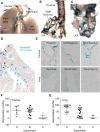
Transplanted mouse TSCs/progenitor cells repopulate the tracheobronchial and intrapulmonary airways. Mouse TSCs were recovered from ROSA26-LacZ transgenic mice, expanded as rim clones, and these “test” cells were instilled into the trachea of Nod/SCID/TCR-γ knockout mice on NA Recovery Day 2. (A) Whole-mount analysis of the trachea and lungs. Test-derived cells are positive for the LacZ protein product β-Gal+ (blue). (B) Whole-mount analysis of the trachea and luminal surface of the right bronchus. (C) Whole-mount analysis of the luminal surface of the left bronchus. (D) Tissue section analysis of the lower right lung lobe. Arrows: dark blue, bronchial region, light blue, bronchiolar region. (E) Section analysis of tracheal and lung subregions. Original magnification, ×200. (F and G) Volume fraction of the tracheal (F) and lung (G) epithelium that is repopulated by transplanted cells. Mean (±SD). Points indicate individual high-power fields. (AC) Distinct host mice transplanted with mouse rim clone cells.
Figure 5.
Figure 5.
Transplanted mouse TSCs/progenitor cells exhibit multilineage differentiation after transplantation. Mice were transplanted as indicated in Figure 4. (AC) Section analysis of the tracheal epithelium: test-derived cells are positive for the LacZ protein product, β-galactosidase (β-Gal)+ (blue). (A) Keratin 5 (brown); (B) CCSP (brown); (C) acetylated tubulin (ACT; brown). (D) Quantification of host-derived (white) and test-derived (blue) basal cells (keratin 5), secretory cells (CCSP), and ciliated cells (ACT). Mean (±SD); n = 3; β-gal (blue). (EH) Section analysis of the intrapulmonary airway epithelium: test-derived cells are β-gal+ (blue). (E and F) CCSP (brown); (G and H) ACT (brown). (E and G) 20×; (F and H) 40×. (I) Quantification of host- (white) and test-derived (blue) basal cells (Keratin 5), secretory cells (CCSP), and ciliated cells (ACT). Mean (±SD); n = 15–50 high-power fields. Arrows signify cell type–specific antigen-positive test cells.
Figure 6.
Figure 6.
Transplanted human TSCs/progenitor cells repopulate the tracheal and intrapulmonary epithelium. Green fluorescent protein–positive human bronchial basal cells were transplanted as indicated in Figure 4. (AC) Immunofluorescence detection of differentiation antigens in the trachea (80×). (D) Quantification of tracheal epithelial cell origin. Minimum/maximum with line at median; n = 3. (E) Quantification of host-derived (H; blue bars) and test-derived (T; green bars) cell distribution in the proximal (Prox) and distal (Dist) trachea. (F) Differentiation of host- and test-derived cells to basal cells (keratin 5), secretory cells (mucin 5B [MUC5B]), or ciliated cells (ACT). Box indicates the range; line indicates the mean; n = 45 high-power fields. (GI) Immunofluorescence detection of differentiation antigens in the intrapulmonary airways (80×). (J) Quantification of intrapulmonary epithelial cell origin. (K) Quantification of epithelial cell origin in airway subregions. BO, bronchiolar; BR, bronchial; H, host; T, test; TB, terminal bronchiolar. Minimum/maximum with line at median; n = 3. (L) Quantification of cell differentiation. Bars indicate the range; box indicates the mean; line indicates the mean; n = 15–26 high-power fields. Arrows signify cell type–specific antigen-positive test cells. DAPI, 4′,6-diamidino-2-phenylindole; GFP, green fluorescent protein.

References

    1. Morrison SJ. Stem cell potential: can anything make anything? Curr Biol. 2001;11:R7–R9. - PubMed
    1. Ghosh M, Smith RW, Runkle CM, Hicks DA, Helm KM, Reynolds SD. Regulation of trachebronchial tissue–specific stem cell pool size. Stem Cells. 2013;31:2767–2778. - PMC - PubMed
    1. Ghosh M, Helm KM, Smith RW, Giordanengo MS, Li B, Shen H, Reynolds SD. A single cell functions as a tissue-specific stem cell and the in vitro niche-forming cell. Am J Respir Cell Mol Biol. 2011;45:459–469. - PMC - PubMed
    1. Hegab AE, Ha VL, Gilbert JL, Zhang KX, Malkoski SP, Chon AT, Darmawan DO, Bisht B, Ooi AT, Pellegrini M, et al. Novel stem/progenitor cell population from murine tracheal submucosal gland ducts with multipotent regenerative potential. Stem Cells. 2011;29:1283–1293. - PMC - PubMed
    1. Hegab AE, Ha VL, Darmawan DO, Gilbert JL, Ooi AT, Attiga YS, Bisht B, Nickerson DW, Gomperts BN. Isolation and in vitro characterization of basal and submucosal gland duct stem/progenitor cells from human proximal airways. Stem Cells Transl Med. 2012;1:719–724. - PMC - PubMed

Publication types