. 2017 Apr;118(4):764-774.
doi: 10.1002/jcb.25736. Epub 2016 Dec 29.

The Ultrastructural Signature of Human Embryonic Stem Cells

Affiliations

The Ultrastructural Signature of Human Embryonic Stem Cells

Jean M Underwood et al. J Cell Biochem. 2017 Apr.

Abstract

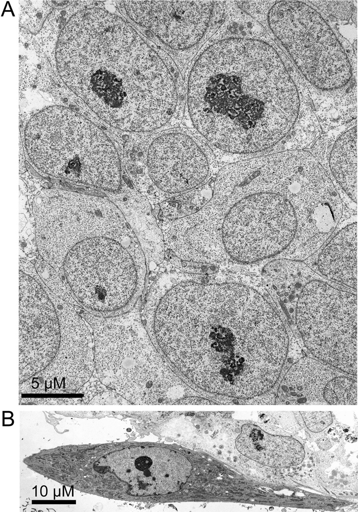
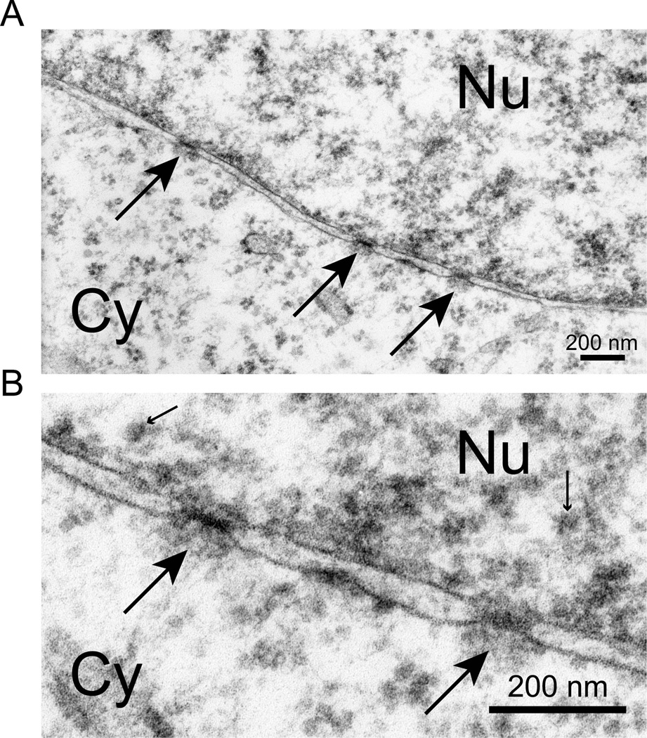
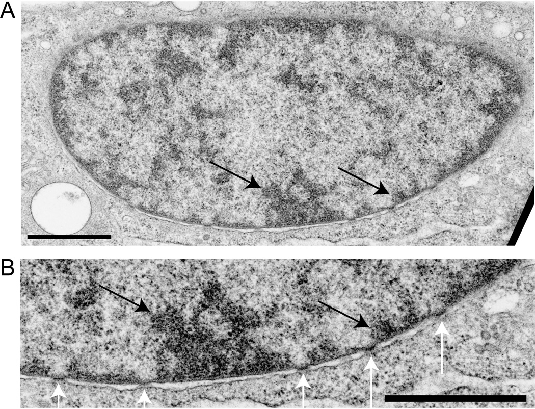
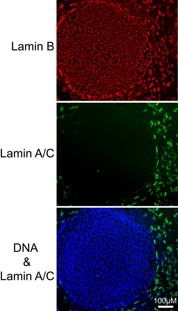
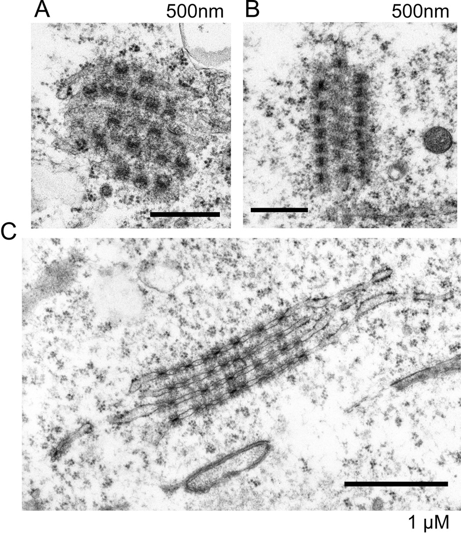
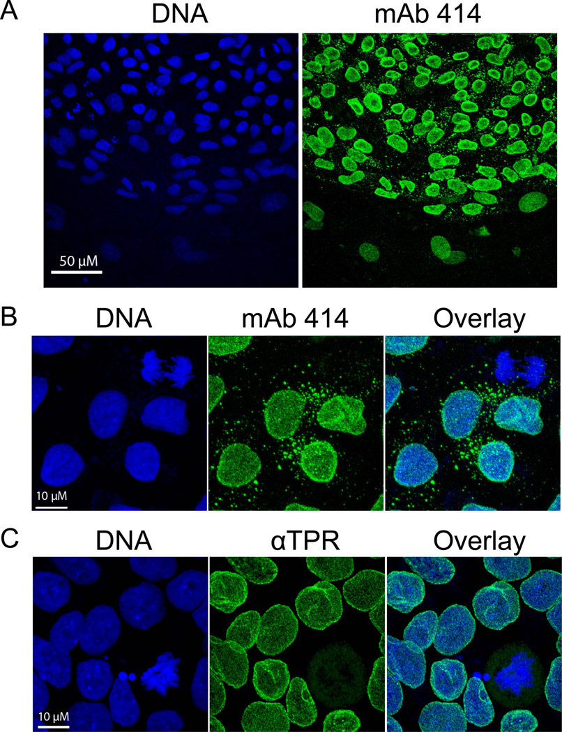
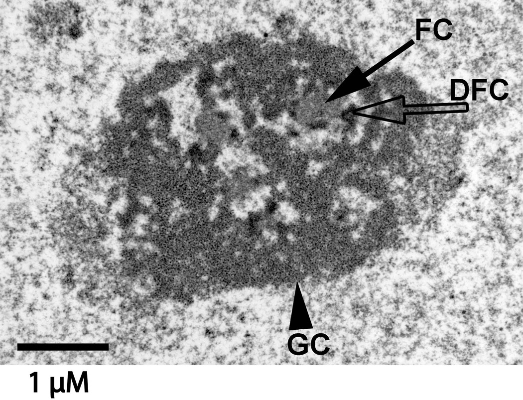
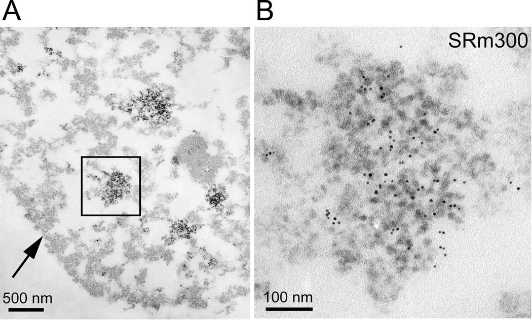
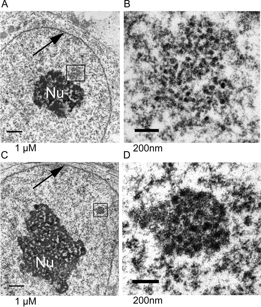
The epigenetics and molecular biology of human embryonic stem cells (hES cells) have received much more attention than their architecture. We present a more complete look at hES cells by electron microscopy, with a special emphasis on the architecture of the nucleus. We propose that there is an ultrastructural signature of pluripotent human cells. hES cell nuclei lack heterochromatin, including the peripheral heterochromatin, that is common in most somatic cell types. The absence of peripheral heterochromatin may be related to the absence of lamins A and C, proteins important for linking chromatin to the nuclear lamina and envelope. Lamins A and C expression and the development of peripheral heterochromatin were early steps in the development of embryoid bodies. While hES cell nuclei had abundant nuclear pores, they also had an abundance of nuclear pores in the cytoplasm in the form of annulate lamellae. These were not a residue of annulate lamellae from germ cells or the early embryos from which hES cells were derived. Subnuclear structures including nucleoli, interchromatin granule clusters, and Cajal bodies were observed in the nuclear interior. The architectural organization of human ES cell nuclei has important implications for cell structure-gene expression relationships and for the maintenance of pluripotency. J. Cell. Biochem. 118: 764-774, 2017. © 2016 Wiley Periodicals, Inc.

Keywords: ANNULATE LAMELLAE; CHROMATIN ORGANIZATION; ELECTRON MICROSCOPY; HUMAN EMBRYONIC STEM CELLS; NUCLEAR STRUCTURE; ULTRASTRUCTURE.


Conflict of interest statement

Disclosure of Potential Conflicts of Interest: The authors have no conflicts to declare.

Figures

Figure 1
Figure 1
A low magnification transmission electron micrograph of H1 hES cells. A colony of H1 cells was fixed and processed as described in Materials and Methods. (A) hES cells grew in tightly packed colonies. Cells were in close contact along all plasma membranes, though cell-cell junctions were not observed. Cells had a high ratio of nuclei to cytoplasm. Cell cytoplasm had an unusually simple ultrastructure, with few inclusions other than mitochondria and lipid droplets that stained slightly after osmium tetroxide postfixation. Nuclei were typically round and also had a simple ultrastructure, usually having a single large nucleolus and a striking absence of heterochromatin. Size bar = 5 µM (B) In contrast, the ultrastructure of a mouse feeder cell at the edge of a hES cell colony was much more complex. The cytoplasm was filled with a much higher density of rough endoplasmic reticulum as well as a higher density of mitochondria. Nuclei had patches of heterochromatin at the nuclear periphery and had the centromeric heterochromatin characteristic of mouse cells. Size bar = 10 µM
Figure 2
Figure 2
hES cells lack heterochromatin. This is a higher magnification view of a H9 hES cell that was in the center of a colony. The cytoplasm is simple with mitochondria (M) and lipid droplets (L) which are stained light grey by the osmium postfixation. As in many cells in this sample, an unusual cytoplasmic structure, an annulate lamellae (AL), was observed. The nucleus (Nu) had a large nucleolus and a striking absence of heterochromatin. Size bar = 2 µM
Figure 3
Figure 3
hES cells have only euchromatin at the interior of the nuclear lamina. This is the edge of an H9 hES cell nucleus shown at two increasing magnifications. The cytoplasm (Cy) has few structures except free polyribosomes. The edge of the nucleus has the two membranes of the nuclear envelope that meet at nuclear pores (large arrows). The nuclear lamina on the nuclear side of the inner nuclear membrane is very thin and barely visible. Chromatin at the inner surface of the lamina was decondensed euchromatin, as in all cells in this study. The dominant feature of this chromatin was small masses of similar size (small arrows), all larger than the ribosomes observed in the cytoplasm. A random selection of these masses was measured as described in Materials and Methods in the micrograph of lower panel as 24.0 µM +/− 1.2µM (n=10), a size consistent with the identification of these as individual nucleosomes coated with the uranyl acetate stain. Electron dense masses of substantially larger size would be present in heterochromatin and these were missing. Both size bars = 200 nM
Figure 4
Figure 4. hES cells develop heterochromatin after differentiation into embryoid bodies
Heterochromatin was observed in almost all cells. Embryoid body formation was induced by withdrawal of FGF and supplementation with serum. These images, at two magnifications, are for induced H9 hES cells at 7 weeks. In Panel B the black arrows mark peripheral heterochromatin and the white arrows show nuclear pores. Size bars = 1 uM
Figure 5
Figure 5. hES cells do not express Lamin A/C
H1 cells grown on a coverslip were simultaneously stained for lamin A/C and Lamin B and imaged by fluorescence microscopy. The large colony of hESC cells stained for Lamin B but not Lamin A/C while the surrounding layer of mouse fibroblast feeder cells contained both Lamins A/C and B in their nuclear laminas. DNA was detected with DAPI.
Figure 6
Figure 6. hESC cells had abundant annulate lamellae
Electron microscopy revealed annulate lamellae in the cytoplasm of many sections for both H1 hES cells (Panel A) and H9 hESCs (Panels B and C). These are three typical views in different orientations. Nuclear pores were embedded in double membranes that were often stacked and were not connected to the nucleus. The size bars are 500nm for Panels A and B and 1 uM for Panel C.
Figure 7
Figure 7. hESC cells had nuclear pores in the cytoplasm that contained FxFG repeat nucleoporins but not TPR
Presented are representative maximum intensity confocal projections of H9 hES cells (Panels A and B) and H1 hESC cells (Panel C). Most nuclear pores were in the nuclear lamina but annulate lamellae were observed as small masses in the cytoplasm of most cells that stained with the MAB414 antibody recognizing nucleoporin p62 and related FxFG nucleoporins [Davis and Blobel, 1986; Sukegawa and Blobel, 1993]. They were not recognized by an antibody specific for TPR, a protein in filamentous projections on the nucleoplasmic face of nuclear pores [Cordes et al., 1997]. DNA was detected by DRAQ5 staining.
Figure 8
Figure 8. The ultrastucture of hES cell nucleoli was not obscured by adjacent heterochromatin
This typical nucleolus was in an H1 hES cell. Subdomains of nucleoli were easily discerned. These were fibrillar centers (FCs), dense fibrillar components (DFCs), and granular components (GC). In one model of nucleolar function [Olson and Dundr, 2005], rDNA transcription takes place at the border of the FC and DFC while rRNA processing begins in the DFC. The granules in the GC are ribosomal subunits. Size bar = 1 uM
Figure 9
Figure 9. hES cells have interchromatin granule clusters
This H9 hES cell was stained with a monoclonal antibody recognizing SRm300 [Blencowe et al., 1998] and a secondary antibody coupled to 5nm gold beads before embedment and sectioning. The EDTA regressive counterstain is selective for RNA [Bernhard, 1969; Monneron and Bernhard, 1969]. Interchromatin granule clusters correspond to the RNA splicing speckled domains seen by fluorescence microscopy and contain high concentrations of RNA processing and export factors. Panel B is a higher magnification view of the interchromatin granule cluster marked in Panel A.
Figure 10
Figure 10. Nuclear bodies observed in hES cells
(A and B) Many nuclei, as in this H9 hES cell, had loose clusters of granules that resembled perichromatin granules [Monneron and Bernhard, 1969]. (C and D) As seen in this H1 hES cell, structures of closely clustered connected granules surrounded by a halo region with a lower concentration of chromatin were observed. These closely resembled the Cajal bodies of somatic cells [Cajal, 1903; Monneron and Bernhard, 1969; Morris, 2008]

References

    1. Ahmed K, Dehghani H, Rugg-Gunn P, Fussner E, Rossant J, Bazett-Jones DP. Global chromatin architecture reflects pluripotency and lineage commitment in the early mouse embryo. PLoS One. 2010;5:e10531. - PMC - PubMed
    1. Andres V, Gonzalez JM. Role of A-type lamins in signaling, transcription, and chromatin organization. J Cell Biol. 2009;187:945–957. - PMC - PubMed
    1. Baricheva EA, Berrios M, Bogachev SS, Borisevich IV, Lapik ER, Sharakhov IV, Stuurman N, Fisher PA. DNA from Drosophila melanogaster beta-heterochromatin binds specifically to nuclear lamins in vitro and the nuclear envelope in situ. Gene. 1996;171:171–176. - PubMed
    1. Bernhard W. A new procedure for electron microscopical cytology. J. Ultrastruct. Res. 1969;27:250–265. - PubMed
    1. Bernstein BE, Mikkelsen TS, Xie X, Kamal M, Huebert DJ, Cuff J, Fry B, Meissner A, Wernig M, Plath K, Jaenisch R, Wagschal A, Feil R, Schreiber SL, Lander ES. A bivalent chromatin structure marks key developmental genes in embryonic stem cells. Cell. 2006;125:315–326. - PubMed

Publication types

MeSH terms

LinkOut - more resources