

TBX18 gene induces adipose-derived stem cells to differentiate into pacemaker-like cells in the myocardial microenvironment
- PMID: 27632938
- PMCID: PMC5065308
- DOI: 10.3892/ijmm.2016.2736
TBX18 gene induces adipose-derived stem cells to differentiate into pacemaker-like cells in the myocardial microenvironment
Abstract
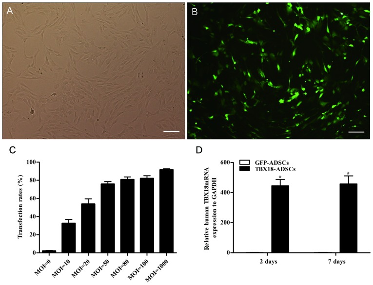
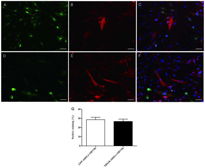
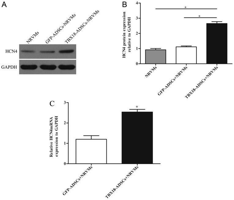
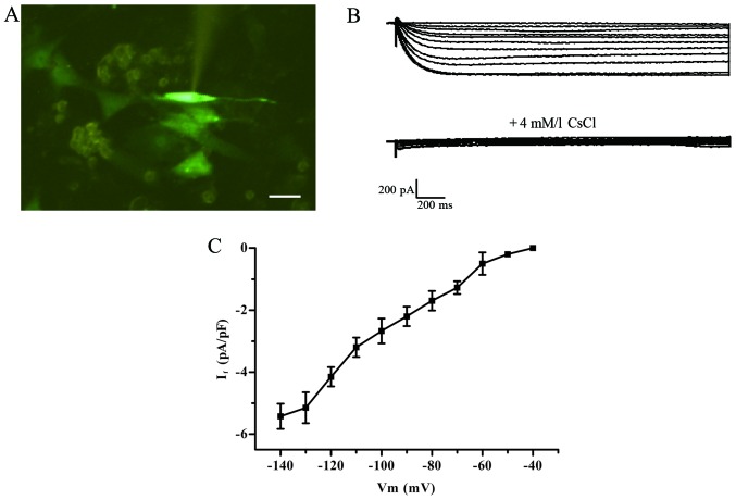
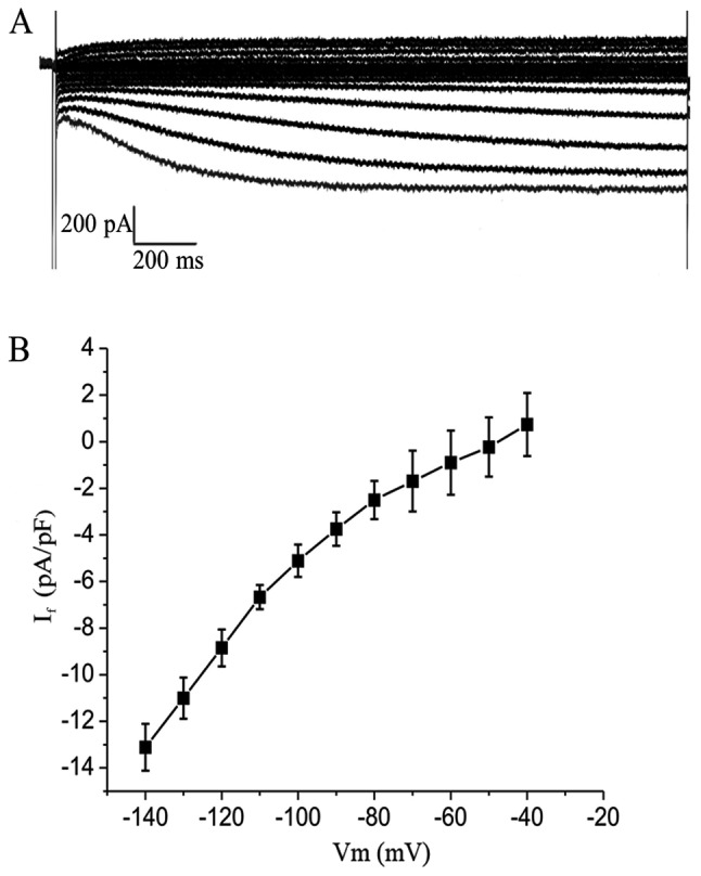
T-box 18 (TBX18) plays a crucial role in the formation and development of the head of the sinoatrial node. The objective of this study was to induce adipose-derived stem cells (ADSCs) to produce pacemaker-like cells by transfection with the TBX18 gene. A recombinant adenovirus vector carrying the human TBX18 gene was constructed to transfect ADSCs. The ADSCs transfected with TBX18 were considered the TBX18-ADSCs. The control group was the GFP-ADSCs. The transfected cells were co-cultured with neonatal rat ventricular cardiomyocytes (NRVMs). The results showed that the mRNA expression of TBX18 in TBX18-ADSCs was significantly higher than in the control group after 48 h and 7 days. After 7 days of co-culturing with NRVMs, there was no significant difference in the expression of the myocardial marker cardiac troponin I (cTnI) between the two groups. RT-qPCR and western blot analysis showed that the expression of HCN4 was higher in the TBX18-ADSCs than in the GFP-ADSCs. The If current was detected using the whole cell patch clamp technique and was blocked by the specific blocker CsCl. Human induced pluripotent stem cell-derived cardiomyocytes (hiPSCMs) showed approximately twice the current density compared with the ADSCs. Our study indicated that the TBX18 gene induces ADSCs to differentiate into pacemaker‑like cells in the cardiac microenvironment. Although further experiments are required in order to assess safety and efficacy prior to implementation in clinical practice, this technique may provide new avenues for the clinical therapy of bradycardia.
Figures
Similar articles
-
TBX18 transcription factor overexpression in human-induced pluripotent stem cells increases their differentiation into pacemaker-like cells.J Cell Physiol. 2019 Feb;234(2):1534-1546. doi: 10.1002/jcp.27018. Epub 2018 Aug 5. J Cell Physiol. 2019. PMID: 30078203
-
Comparison of mouse brown and white adipose‑derived stem cell differentiation into pacemaker‑like cells induced by TBX18 transduction.Mol Med Rep. 2018 May;17(5):7055-7064. doi: 10.3892/mmr.2018.8792. Epub 2018 Mar 20. Mol Med Rep. 2018. PMID: 29568953 Free PMC article.
-
Transcription factor TBX18 promotes adult rat bone mesenchymal stem cell differentiation to biological pacemaker cells.Int J Mol Med. 2018 Feb;41(2):845-851. doi: 10.3892/ijmm.2017.3259. Epub 2017 Nov 16. Int J Mol Med. 2018. PMID: 29207072 Free PMC article.
-
[Electrophysiological properties of stem cells].Herz. 2006 Apr;31(2):123-6. doi: 10.1007/s00059-006-2793-y. Herz. 2006. PMID: 16738835 Review. German.
-
Harvesting, Isolation and Differentiation of Rat Adipose-Derived Stem Cells.Curr Pharm Biotechnol. 2018;19(1):19-29. doi: 10.2174/1389201019666180418101323. Curr Pharm Biotechnol. 2018. PMID: 29667552 Review.
Cited by
-
Tbx18-dependent differentiation of brown adipose tissue-derived stem cells toward cardiac pacemaker cells.Mol Cell Biochem. 2017 Sep;433(1-2):61-77. doi: 10.1007/s11010-017-3016-y. Epub 2017 Apr 5. Mol Cell Biochem. 2017. PMID: 28382491
-
Conversion of Unmodified Stem Cells to Pacemaker Cells by Overexpression of Key Developmental Genes.Cells. 2023 May 13;12(10):1381. doi: 10.3390/cells12101381. Cells. 2023. PMID: 37408215 Free PMC article.
-
Enrichment differentiation of human induced pluripotent stem cells into sinoatrial node-like cells by combined modulation of BMP, FGF, and RA signaling pathways.Stem Cell Res Ther. 2020 Jul 16;11(1):284. doi: 10.1186/s13287-020-01794-5. Stem Cell Res Ther. 2020. PMID: 32678003 Free PMC article.
-
Plasticity of Adipose Tissue-Derived Stem Cells and Regulation of Angiogenesis.Front Physiol. 2018 Nov 26;9:1656. doi: 10.3389/fphys.2018.01656. eCollection 2018. Front Physiol. 2018. PMID: 30534080 Free PMC article. Review.
-
Reciprocal interaction between IK1 and If in biological pacemakers: A simulation study.PLoS Comput Biol. 2021 Mar 10;17(3):e1008177. doi: 10.1371/journal.pcbi.1008177. eCollection 2021 Mar. PLoS Comput Biol. 2021. PMID: 33690622 Free PMC article.
References
-
- Bai X, Yan Y, Song YH, Seidensticker M, Rabinovich B, Metzele R, Bankson JA, Vykoukal D, Alt E. Both cultured and freshly isolated adipose tissue-derived stem cells enhance cardiac function after acute myocardial infarction. Eur Heart J. 2010;31:489–501. doi: 10.1093/eurheartj/ehp568. - DOI - PubMed
MeSH terms
Substances
LinkOut - more resources
Full Text Sources
Other Literature Sources
Medical
Research Materials
