. 2016 Sep 1;30(17):1937-42.
doi: 10.1101/gad.285767.116. Epub 2016 Sep 15.

Polarized regulatory landscape and Wnt responsiveness underlie Hox activation in embryos

Affiliations

Polarized regulatory landscape and Wnt responsiveness underlie Hox activation in embryos

Roel Neijts et al. Genes Dev. .

Abstract

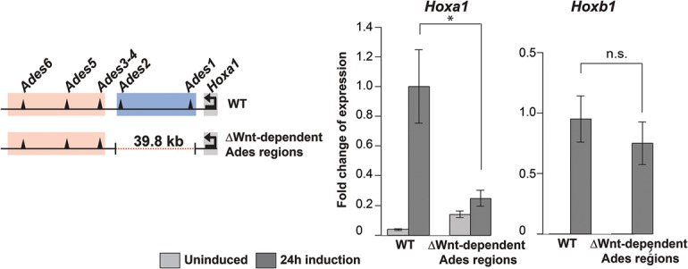
Sequential 3'-to-5' activation of the Hox gene clusters in early embryos is a most fascinating issue in developmental biology. Neither the trigger nor the regulatory elements involved in the transcriptional initiation of the 3'-most Hox genes have been unraveled in any organism. We demonstrate that a series of enhancers, some of which are Wnt-dependent, is located within a HoxA 3' subtopologically associated domain (subTAD). This subTAD forms the structural basis for multiple layers of 3'-polarized features, including DNA accessibility and enhancer activation. Deletion of the cassette of Wnt-dependent enhancers proves its crucial role in initial transcription of HoxA at the 3' side of the cluster.

Keywords: DNA accessibility; Hox regulation; chromatin conformation; developmental enhancers; regulatory landscapes.


Figures

Figure 1.
Figure 1.
Transcriptional initiation of HoxA in early embryos and in epiblast stem cells (EpiSCs). (A) Early expression pattern of Hoxa1 in gastrulating embryos. (A) Anterior; (P) posterior. (B) Precocious induction of Hoxa1 expression in an E6.0 embryo by Chiron (10 h). Bar, 100 µm. (C) Parallelism between induction of Hoxa1 in embryos and Wnt-stimulated induction of Hoxa1 in EpiSCs. (IWP2) Wnt inhibitor. (D) Kinetics of induction of HoxA genes by Chiron in wild-type EpiSCs. Transcription measured by RT-qPCR is relative to the highest value of expression. Error bars indicate ±SD. (*) P < 0.05; (**) P < 0.01. (E) H3K27me3 and H3K4me3 marks along the HoxA cluster in uninduced (0 h) and Wnt-induced (12 and 72 h) Wnt3-null EpiSCs.
Figure 2.
Figure 2.
Interactions between the HoxA locus and putative Ades enhancers. (A) Zoom in on 3′ subTAD region with 4C-seq profile from the Hoxa1 viewpoint (red dotted line) and distribution of H3K27ac in uninduced (0 h) and Chiron-induced (24 h) EpiSCs. Positions acetylated before induction (Ades3–4, Ades5, and Ades6) and positions becoming acetylated after induction (Ades1 and Ades2) are highlighted in red and blue, respectively. β-Catenin (β-cat)-binding regions (Zhang et al. 2013) are indicated. (B) 4C-seq profiles from Ades enhancers and HoxA viewpoints in uninduced (−) and Wnt-stimulated (+) conditions. The patterns of interactions define a proximal and a distal subpart of the 3′ subTAD. Wnt stimulation results in more compaction of these segments (arrows). The HoxA cluster appears to comprise three parts, indicated below. See also Supplemental Figures S5 and S6.
Figure 3.
Figure 3.
Activity and DNA accessibility of the Ades enhancers. (A, top row) Activity of Ades enhancers coupled to lacZ in E7.5 to E7.8 (head fold to early somite) embryos. (Right) Hoxa1 expression. (Bottom row) The earliest embryonic stage at which each enhancer is observed to be active (varying from E6.5 [before actual Hoxa1 expression] to E7.5). (Black curved line) Region of activity; (dotted line) boundary between embryonic (below the line) and extraembryonic (above the line) tissues. (B) ATAC-seq (assay for transposase-accessible chromatin [ATAC] with high-throughput sequencing) profile of EpiSCs (uninduced and after 48 h of Chiron activation) and in pre-Hox (E6.0), early Hox (E7.2), and later Hox (posterior tissues of E7.8) embryos in the Ades region. Bars, 100 µm.
Figure 4.
Figure 4.
The most proximal region of the 3′ subTAD is required to activate Hoxa1. Deletion of the Wnt-dependent Ades1 and Ades2 region reduces Hoxa1 transcriptional response to Chiron (24 h); Hoxb1 is unaffected. Transcription measured by RT-qPCR is relative to the highest value of expression. Errors bars indicate ±SD. (*) P < 0.05.
Figure 5.
Figure 5.
Model summarizing the findings of distinct steps leading to transcriptional initiation of HoxA genes. Three successive phases of 3′-oriented epigenetic events culminate in 3′ Hox gene transcription. (1) Tropism of contacts between 3′ HoxA and the 3′ surrounding in ESCs (Hox ground state) and its compaction to the 3′ subTAD in EpiSCs (preprimed Hox state). (2) Accessibility of Wnt-dependent proximal elements (open triangles) in the proximal 3′ subTAD appears between uninduced EpiSCs and E6.0 embryos. (3) Acetylation of these enhancers and 3′ HoxA transcription arise at E7.2. At that stage, all enhancers are acetylated. More 5′ HoxA genes are subsequently expressed (E7.8).

References

    1. Barrow JR, Thomas KR, Boussadia-Zahui O, Moore R, Kemler R, Capecchi MR, McMahon AP. 2003. Ectodermal Wnt3/β-catenin signaling is required for the establishment and maintenance of the apical ectodermal ridge. Genes Dev 17: 394–409. - PMC - PubMed
    1. Berlivet S, Paquette D, Dumouchel A, Langlais D, Dostie J, Kmita M. 2013. Clustering of tissue-specific sub-TADs accompanies the regulation of HoxA genes in developing limbs. PLoS Genet 9: e1004018. - PMC - PubMed
    1. Brons IG, Smithers LE, Trotter MW, Rugg-Gunn P, Sun B, Chuva de Sousa Lopes SM, Howlett SK, Clarkson A, Ahrlund-Richter L, Pedersen RA, et al. 2007. Derivation of pluripotent epiblast stem cells from mammalian embryos. Nature 448: 191–195. - PubMed
    1. Buenrostro JD, Giresi PG, Zaba LC, Chang HY, Greenleaf WJ. 2013. Transposition of native chromatin for fast and sensitive epigenomic profiling of open chromatin, DNA-binding proteins and nucleosome position. Nat Methods 10: 1213–1218. - PMC - PubMed
    1. Cong L, Ran FA, Cox D, Lin S, Barretto R, Habib N, Hsu PD, Wu X, Jiang W, Marraffini LA, et al. 2013. Multiplex genome engineering using CRISPR/Cas systems. Science 339: 819–823. - PMC - PubMed