Review
. 2016 Sep 16;11(1):99.
doi: 10.1186/s13018-016-0432-8.

Sports-related wrist and hand injuries: a review

Affiliations
Review

Sports-related wrist and hand injuries: a review

Daniel M Avery 3rd et al. J Orthop Surg Res. .

Abstract

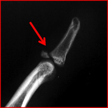
Background: Hand and wrist injuries are common during athletics and can have a significant impact especially if initially disregarded. Due to their high level of physical demand, athletes represent a unique subset of the population.

Main body: The following is an overview of hand and wrist injuries commonly seen in athletics. Information regarding evaluation, diagnosis, conservative measures, and surgical treatment are provided.

Conclusion: Knowledge of these entities and special consideration for the athlete can help the team physician effectively treat these players and help them achieve their goals.

Keywords: Fracture; Hand injuries; Ligament; Return to play; Sports; Surgical treatment; Wrist injuries.


Figures

Fig. 1
Fig. 1
a PA radiograph of a nondisplaced proximal pole scaphoid fracture in a recreational hockey player. b PA radiograph of a nondisplaced scaphoid waist fracture in a high school soccer player treated with headless compression screw fixation
Fig. 2
Fig. 2
a PA radiograph showing a flexed scaphoid (signet ring sign). b Intraoperative finding of a complete SL interosseous ligament tear with the tip of the probe on the scaphoid (yellow arrow). c Open reduction of the SL interval (blue arrow) prior to ligament repair
Fig. 3
Fig. 3
a Intraoperative finding of a volarly subluxed ECU tendon (between yellow lines) in a recreational tennis player. b The ECU tendon back in its reduced position (red lines) after an anatomic repair of the subsheath
Fig. 4
Fig. 4
a PA radiograph of a patient with ulnar abutment revealing both 6 mm of ulnar-positive variance and incidentally an ulnar styloid nonunion. b Neutral to −1 mm of ulnar variance after a diaphyseal ulnar shortening osteotomy is performed
Fig. 5
Fig. 5
a A T2-weighted coronal sequence of a wrist MRI revealing an ulnar-sided peripheral tear of the TFCC (yellow arrow). b Arthroscopy view from the three to four portal showing the peripheral tear (red arrow). c Intraoperative arthrosopic image during an arthroscopic-assisted outside-in repair using PDS suture (blue arrow)
Fig. 6
Fig. 6
Axial CT image demonstrating a hook of the hamate fracture (red arrow) in a college hockey player
Fig. 7
Fig. 7
a Preoperative photograph demonstrating a patient with no endpoint to valgus stress testing of the thumb MCP joint. b A T2-weighted coronal sequence demonstrating a complete tear of the UCL which is detached from the proximal phalanx (yellow arrow)
Fig. 8
Fig. 8
a Intraoperative and b fluoroscopic images of a long oblique metacarpal shaft fracture secured with three lag screws. c Intraoperative and d fluoroscopic images of a transverse shaft fracture secured with a plate and screws
Fig. 9
Fig. 9
Plate and screw fixation of an angulated fifth metacarpal neck fracture in a high school running back
Fig. 10
Fig. 10
A PA radiograph of a small finger transverse proximal phalanx fracture with clinical malrotation which was treated with closed reduction and crossed K-wires
Fig. 11
Fig. 11
Lateral radiograph of a dorsal fracture dislocation of the PIP joint with 50 % articular involvement of the middle phalanx
Fig. 12
Fig. 12
a Intraoperative findings of a patient with a torn radial sagittal band and ulnarly subluxed EDC tendon before repair b and after repair
Fig. 13
Fig. 13
Lateral radiograph of a ring finger of a patient whose finger got caught on a basketball net showing an FDP bony avulsion fracture with the fragment caught up distal to the A4 pulley level (yellow arrow)
Fig. 14
Fig. 14
Lateral radiograph of a mallet finger with a bony avulsion fragment (red arrow)

References

    1. Rettig AC. Athletic injuries of the wrist and hand. Part 1. Traumatic injuries of the wrist. Am J Sports Med. 2003;31:1038–48. - PubMed
    1. Amadio PC. Epidemiology of hand and wrist injuries in sports. Hand Clin. 1990;6:379–81. - PubMed
    1. Geissler WB, Burkett JL. Ligamentous sports injuries of the hand and wrist. Sports Med Arthrosc Rev. 2014;22(1):39–44. doi: 10.1097/JSA.0000000000000013. - DOI - PubMed
    1. Sendher R, Ladd AL. The scaphoid. Orthop Clin North Am. 2013;44(1):107–20. doi: 10.1016/j.ocl.2012.09.003. - DOI - PubMed
    1. Geissler WB. Arthroscopic management of scaphoid fractures in athletes. Hand Clin. 2009;25(3):359–69. doi: 10.1016/j.hcl.2009.05.004. - DOI - PubMed

MeSH terms