

Characterization and mechanisms of the pharyngeal swallow activated by stimulation of the esophagus
- PMID: 27634013
- PMCID: PMC5130554
- DOI: 10.1152/ajpgi.00291.2016
Characterization and mechanisms of the pharyngeal swallow activated by stimulation of the esophagus
Abstract
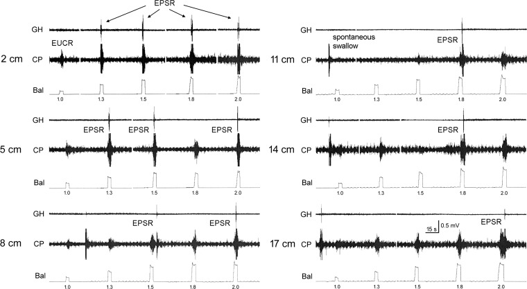
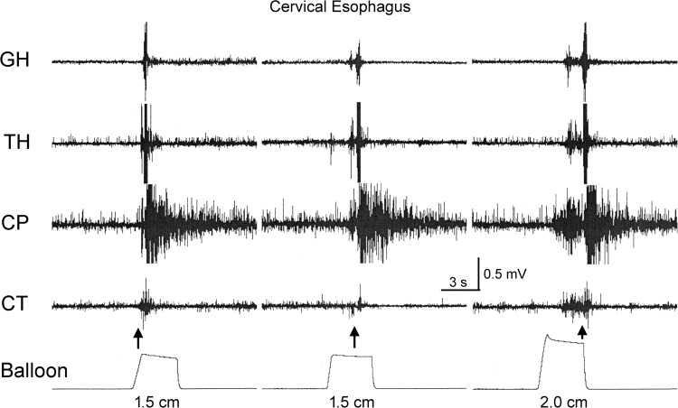
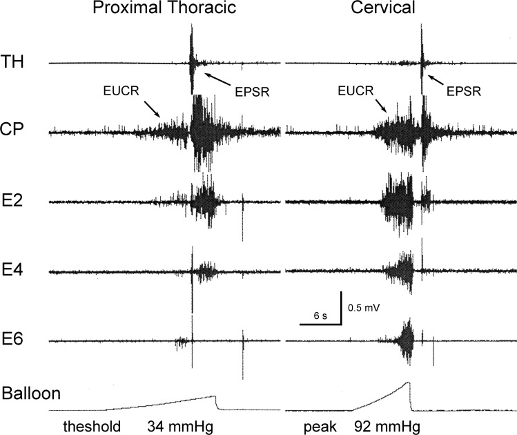
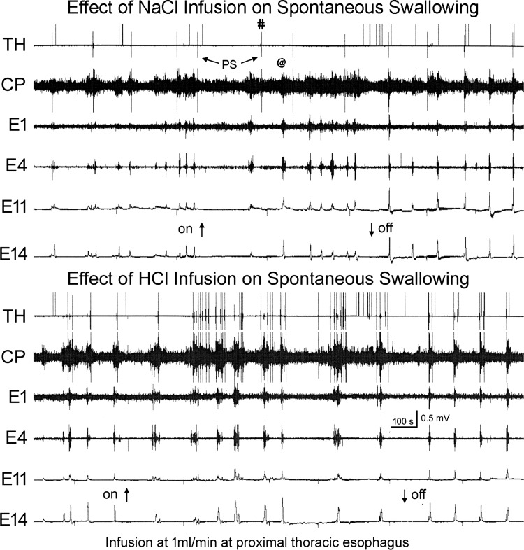
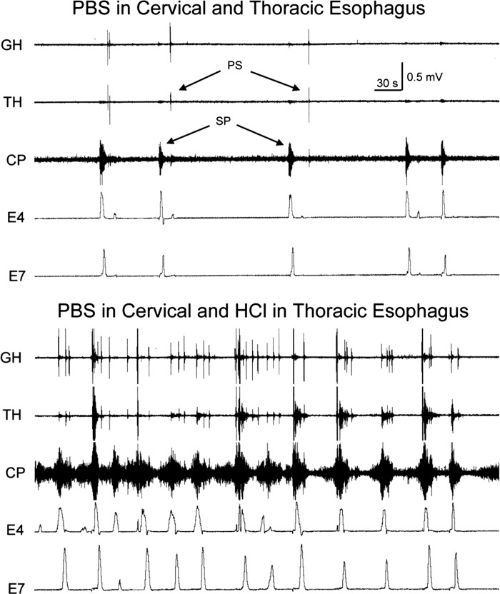
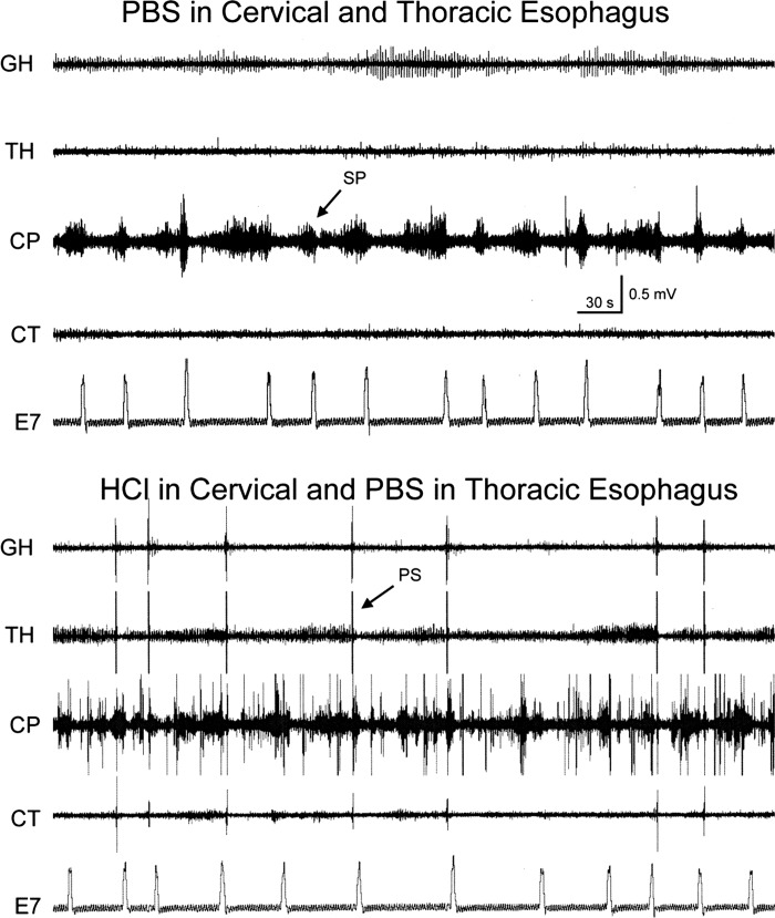
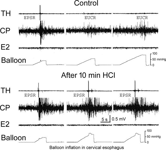
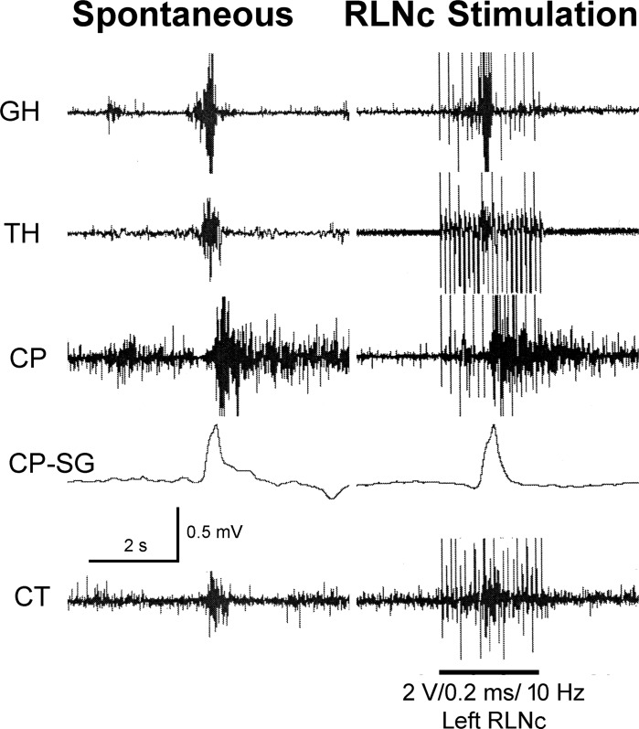
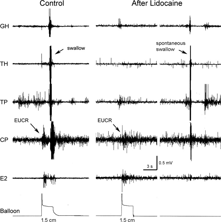
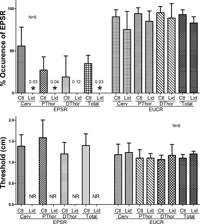
Stimulation of the esophagus activates the pharyngeal swallow response (EPSR) in human infants and animals. The aims of this study were to characterize the stimulus and response of the EPSR and to determine the function and mechanisms generating the EPSR. Studies were conducted in 46 decerebrate cats in which pharyngeal, laryngeal, and esophageal motility was monitored using EMG, strain gauges, or manometry. The esophagus was stimulated by balloon distension or luminal fluid infusion. We found that esophageal distension increased the chance of occurrence of the EPSR, but the delay was variable. The chance of occurrence of the EPSR was related to the position, magnitude, and length of the stimulus in the esophagus. The most effective stimulus was long, strong, and situated in the cervical esophagus. Acidification of the esophagus activated pharyngeal swallows and sensitized the receptors that activate the EPSR. The EPSR was blocked by local anesthesia applied to the esophageal lumen, and electrical stimulation of the recurrent laryngeal nerve caudal to the cricoid cartilage (RLNc) activated the pharyngeal swallow response. We conclude that the EPSR is activated in a probabilistic manner. The receptors mediating the EPSR are probably mucosal slowly adapting tension receptors. The sensory neural pathway includes the RLNc and superior laryngeal nerve. We hypothesize that, because the EPSR is observed in human infants and animals, but not human adults, activation of EPSR is related to the elevated position of the larynx. In this situation, the EPSR occurs rather than secondary peristalsis to prevent supraesophageal reflux when the esophageal bolus is in the proximal esophagus.
Keywords: esophagus; pharyngeal swallow; superior laryngeal nerve.
Copyright © 2016 the American Physiological Society.
Figures
Similar articles
-
The effect of body position on esophageal reflexes in cats: a possible mechanism of SIDS?Pediatr Res. 2018 Mar;83(3):731-738. doi: 10.1038/pr.2017.302. Epub 2017 Dec 20. Pediatr Res. 2018. PMID: 29166377 Free PMC article.
-
The role of the superior laryngeal nerve in esophageal reflexes.Am J Physiol Gastrointest Liver Physiol. 2012 Jun 15;302(12):G1445-57. doi: 10.1152/ajpgi.00007.2012. Epub 2012 Mar 8. Am J Physiol Gastrointest Liver Physiol. 2012. PMID: 22403790 Free PMC article.
-
Biomechanical increase in cervical esophageal wall tension during peristalsis.Am J Physiol Gastrointest Liver Physiol. 2024 Jun 1;326(6):G726-G735. doi: 10.1152/ajpgi.00049.2024. Epub 2024 Apr 16. Am J Physiol Gastrointest Liver Physiol. 2024. PMID: 38626405 Free PMC article.
-
Coordination of peristalsis in pharynx and esophagus.Dysphagia. 1993;8(2):74-8. doi: 10.1007/BF02266983. Dysphagia. 1993. PMID: 8467727 Review.
-
Reflex-mediated enhancement of airway protective mechanisms.Am J Med. 2000 Mar 6;108 Suppl 4a:8S-14S. doi: 10.1016/s0002-9343(99)00289-2. Am J Med. 2000. PMID: 10718445 Review.
Cited by
-
Vagus Nerves Provide a Robust Afferent Innervation of the Mucosa Throughout the Body of the Esophagus in the Mouse.Dysphagia. 2020 Jun;35(3):471-478. doi: 10.1007/s00455-019-10051-8. Epub 2019 Aug 29. Dysphagia. 2020. PMID: 31468191 Free PMC article.
-
Serotonin therapies for opioid-induced disordered swallow and respiratory depression.J Appl Physiol (1985). 2024 Apr 1;136(4):821-843. doi: 10.1152/japplphysiol.00509.2023. Epub 2024 Feb 22. J Appl Physiol (1985). 2024. PMID: 38385184 Free PMC article.
-
The effects of simulated gastroesophageal reflux on infant pig oropharyngeal feeding physiology.Am J Physiol Gastrointest Liver Physiol. 2024 Jul 1;327(1):G105-G116. doi: 10.1152/ajpgi.00027.2024. Epub 2024 May 21. Am J Physiol Gastrointest Liver Physiol. 2024. PMID: 38772905 Free PMC article.
-
A biomechanical response of the esophagus participates in swallowing.Am J Physiol Gastrointest Liver Physiol. 2023 Feb 1;324(2):G131-G141. doi: 10.1152/ajpgi.00219.2022. Epub 2022 Dec 13. Am J Physiol Gastrointest Liver Physiol. 2023. PMID: 36511513 Free PMC article.
-
The effect of body position on esophageal reflexes in cats: a possible mechanism of SIDS?Pediatr Res. 2018 Mar;83(3):731-738. doi: 10.1038/pr.2017.302. Epub 2017 Dec 20. Pediatr Res. 2018. PMID: 29166377 Free PMC article.
References
-
- Al-Adnani M, Cohen MC, Scheinberg I. Gastroesophageal reflux disease and sudden infant death syndrome: Mechanisms behind an under-recognized association. Pediatr Dev Pathol 14: 53–56, 2011. - PubMed
-
- Amarasena J, Ootaki S, Yamamura K, Yamada Y. Effect of cortical masticatory area stimulation on swallowing on anesthetized rabbits. Brain Res 965: 222–238, 2003. - PubMed
-
- Aziz Q, Rothwell JC, Barlow J, Thompson DG. Modulation of esophageal responses to magnetic stimulation of the human brain by swallowing and by vagal stimulation. Gastroenterology 109: 1437–1445, 1995. - PubMed
-
- Christensen J. Patterns and origin of some esophageal responses to stretch and electrical stimulation. Gastroenterology 59: 909–916, 1970. - PubMed
Publication types
MeSH terms
Grants and funding
LinkOut - more resources
Full Text Sources
Other Literature Sources
Miscellaneous