. 2016 Dec 1;76(23):6901-6910.
doi: 10.1158/0008-5472.CAN-16-0517. Epub 2016 Sep 7.

Cell Adhesion Molecule CD166 Drives Malignant Progression and Osteolytic Disease in Multiple Myeloma

Affiliations

Cell Adhesion Molecule CD166 Drives Malignant Progression and Osteolytic Disease in Multiple Myeloma

Linlin Xu et al. Cancer Res. .

Abstract

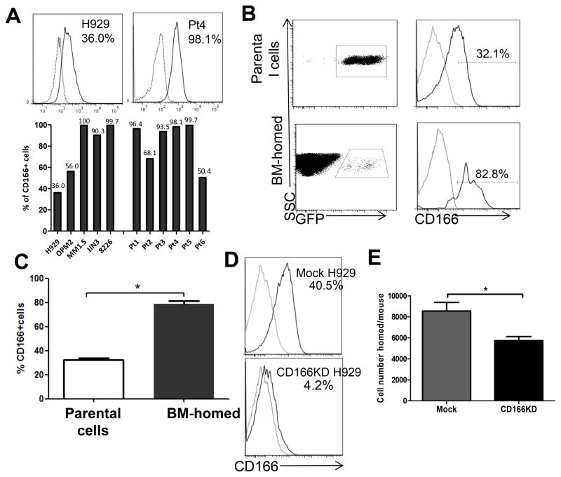
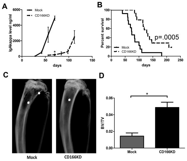
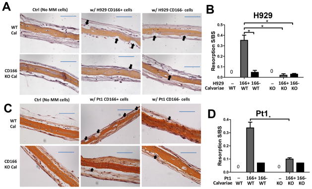
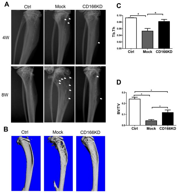
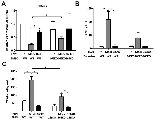
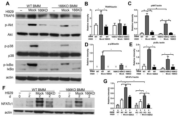
Multiple myeloma is incurable once osteolytic lesions have seeded at skeletal sites, but factors mediating this deadly pathogenic advance remain poorly understood. Here, we report evidence of a major role for the cell adhesion molecule CD166, which we discovered to be highly expressed in multiple myeloma cell lines and primary bone marrow cells from patients. CD166+ multiple myeloma cells homed more efficiently than CD166- cells to the bone marrow of engrafted immunodeficient NSG mice. CD166 silencing in multiple myeloma cells enabled longer survival, a smaller tumor burden, and less osteolytic lesions, as compared with mice bearing control cells. CD166 deficiency in multiple myeloma cell lines or CD138+ bone marrow cells from multiple myeloma patients compromised their ability to induce bone resorption in an ex vivo organ culture system. Furthermore, CD166 deficiency in multiple myeloma cells also reduced the formation of osteolytic disease in vivo after intratibial engraftment. Mechanistic investigation revealed that CD166 expression in multiple myeloma cells inhibited osteoblastogenesis of bone marrow-derived osteoblast progenitors by suppressing Runx2 gene expression. Conversely, CD166 expression in multiple myeloma cells promoted osteoclastogenesis by activating TRAF6-dependent signaling pathways in osteoclast progenitors. Overall, our results define CD166 as a pivotal director in multiple myeloma cell homing to the bone marrow and multiple myeloma progression, rationalizing its further study as a candidate therapeutic target for multiple myeloma treatment. Cancer Res; 76(23); 6901-10. ©2016 AACR.


Conflict of interest statement

The authors declare no conflict of interest.

Figures

Figure 1
Figure 1. CD166 is expressed in both MM cell lines and primary MM CD138+ cells and is critical for homing of MM cells to the BM of NSG mice
(A) Representative flow cytometric analysis of CD166 expression level on H929 cells and MM patients’ CD138+ cells (upper panel). The levels of CD166 expression on MM cell lines and 6 MM patients’ CD138+ cells (lower panel). (B–C) A total of 2×10⁷ GFP-labeled H929 cells were intravenously injected into sub-lethally irradiated NSG mice and GFP+ cells were recovered from mice BM 14h later. The percentage of CD166+ cells within parental H929 cells and from BM-homed cells were compared flow cytometrically. Data are represented as mean ± SEM from 3 pooled experiments (N = 3 mice/group/experiment, each assayed individually). (D) Flow cytometric assessment of the level of CD166 knockdown with lentiviral shRNA for huCD166. (E) 2×10⁷ GFP-labeled mock or CD166KD H929 cells were intravenously injected into sub-lethally irradiated NSG mice. GFP cells were recovered from mouse BM 14h later and the number of H929 cells homed to the BM from two femurs and two tibiae was calculated. Data are represented as mean ± SEM from 3 pooled experiments (N = 3 mice/group/experiment, each assayed individually). *p<0.05. For histograms in A and D: grey line = isotype control, black line = sample.
Figure 2
Figure 2. CD166 is critical for MM disease progression
(A&B) 1×10⁵ Mock or CD166KD H929 cells were intravenously injected into NSG mice. (A) Human IgA-kappa levels in the serum of individual mice were measured by ELISA every two weeks. (B) Survival was monitored for a period of 220 days and Kaplan-Meier survival curves were plotted. Data are pooled from 2 independent experiments (N = 6–8 mice/group/experiment were followed until day 220 post-inoculation). (C) Eight weeks after inoculation, some of the mice were euthanized. Tibiae were imaged with radiography and representative images are shown. Bone lesions area were indicated with white arrows. (D) Eight weeks after inoculation, some of the mice were euthanized. Tibiae were analyzed with micro-CT and trabecular bone volume (BV/TV fraction) were analyzed by an analyzer blinded to the experimental groups. Data are representative of 2 separate experiments (mean±SEM, N = 6 mice/group/experiment, each assayed individually). *p<0.05.
Figure 3
Figure 3. Absence of CD166 on myeloma cells reduces bone resorption on calvariae ex vivo
(A–D) Ex vivo organ culture assay (EVOCA) was used to examine the effect of CD166 expression on myeloma bone resorption lesions. After culture of MM cells on calvariae as described in Methods, calvariae were fixed, decalcified, sectioned and processed for H&E staining. Bone resorption (black arrow) on calvariae was analyzed from three non-overlapping fields per bone under 20 x magnification. The quantitative representation of EVOCA assay was performed by measuring and calculating resorption surface to bone surface (BS) ratio with Bioquant software 2014. (A) H&E staining of Calvariae from WT or CD166KO pups cultured with flow sorted 2×10⁴ CD166+ or CD166− H929 cells for 10 days (×20, scale bar=100 μm). (B) Quantitative representation of EVOCA results with H929; Data represent 3 separate experiments done in triplicates for each group and are expressed as mean± SEM, *p<0.05. (C) H&E staining of calvariae from WT or CD166KO pups cultured with 2×10⁴ flow cytometrically sorted CD166+ or CD166− primary MM CD138+ cells for 10 days (×20, scale bar=100 μm). (D) Quantitative representation of EVOCA results shown in (C).
Figure 4
Figure 4. Absence of CD166 on MM cell leads to less bone osteolytic lesions
(A) Representative radiographic images of tibiae from mice inoculated with mock or CD166KD H929 cells at 4 weeks and 8 weeks after inoculation. Bone lesion areas are indicated with white arrow. (B) Nine weeks after inoculation, mice were euthanized and tibiae were collected and scanned using micro-CT for 3D reconstruction. Bones from 3 representative mice are shown. (C) Trabecular thickness Tb.Th, and (D) trabecular bone volume (BV/TV fraction) were determined by micro-CT readings. n=5–6/group, mean± SEM, *p<0.05.
Figure 5
Figure 5. Expression of CD166 on myeloma cells alters bone remodeling balance
(A) BMSC were cocultured with H929 cells (as indicated) for 48h. H929 cells were removed and RNA was isolated from BMSC. Relative expression of RUNX2 RNA in BMSC was detected by quantitative PCR relative to GAPDH and the “WT BMSC alone” sample. (B) Calvariae were cocultured with H929 cells (as indicated) for 7 days. H929 cells were removed. Relative RANKL and OPG RNA expression in calvariae cells were examined by quantitative PCR and RANKL/OPG ratio was calculated. (C) Non-adherent BM derived macrophage from WT or CD166KO mice were cultured for 3 days to enrich for adherent BM macrophages (BMM). BMM were then cultured in α-MEM in the presence of recombinant mouse M-CSF (10 ng/ml) and RANKL (50ng/ml) for 7 days and mock H929 or CD166KD H929, followed by TRAP staining. TRAP-positive cells with 3 or more nuclei were scored under an inverted microscope. Data represent 3 separate experiments done in triplicates for each group and are expressed as mean± SEM, *p<0.05.
Figure 6
Figure 6. Absence of CD166 expression on MM cells downregulates key signaling pathways in osteoclastogenesis
BMM were derived from bone marrow cells by culturing in the presence of M-SCF (10 ng/ml) for 3 days. (A) BMM were serum starved for 2h before exposure to mock H929 or CD166KD H929 for 30 min. Protein was extracted from BMM by RIPA lysis buffer for Western Blot analyses after H929 cells were washed off with cold PBS. Whole cell extracts were subjected to Western blot analysis with specific Abs as indicated. (F) BMM were cultured with mock H929 or CD166KD H929 in the presence of M-CSF (10 ng/ml) and RANKL (50ng/ml) for the 2 days. Protein was extracted from BMM by RIPA lysis buffer for Western Blot analyses after H929 cells were washed off with cold PBS. Whole cell extracts were subjected to Western blot analysis with specific Abs as indicated. (B–E, G) Quantitative densitometry of the expression of the indicated proteins was normalized to actin protein expression. Data were collected from three separate observations and are expressed as mean± SEM. *p<0.05.

References

    1. Slovak ML. Multiple myeloma: current perspectives. Clinics in laboratory medicine. 2011;31(4):699–724. x. - PubMed
    1. Mitsiades CS, McMillin DW, Klippel S, Hideshima T, Chauhan D, Richardson PG, et al. The role of the bone marrow microenvironment in the pathophysiology of myeloma and its significance in the development of more effective therapies. Hematology/oncology clinics of North America. 2007;21(6):1007–34. vii–viii. - PubMed
    1. Podar K, Chauhan D, Anderson KC. Bone marrow microenvironment and the identification of new targets for myeloma therapy. Leukemia: official journal of the Leukemia Society of America, Leukemia Research Fund, UK. 2009;23(1):10–24. - PMC - PubMed
    1. Van Camp B, Van Riet I. Homing mechanisms in the biology of multiple myeloma. Verhandelingen - Koninklijke Academie voor Geneeskunde van Belgie. 1998;60(3):163–94. - PubMed
    1. Alsayed Y, Ngo H, Runnels J, Leleu X, Singha UK, Pitsillides CM, et al. Mechanisms of regulation of CXCR4/SDF-1 (CXCL12)-dependent migration and homing in multiple myeloma. Blood. 2007;109(7):2708–17. - PMC - PubMed

Publication types