Randomized Controlled Trial
. 2016 Nov 1;39(11):1973-1983.
doi: 10.5665/sleep.6226.

The Combination of Supplemental Oxygen and a Hypnotic Markedly Improves Obstructive Sleep Apnea in Patients with a Mild to Moderate Upper Airway Collapsibility

Affiliations
Randomized Controlled Trial

The Combination of Supplemental Oxygen and a Hypnotic Markedly Improves Obstructive Sleep Apnea in Patients with a Mild to Moderate Upper Airway Collapsibility

Bradley A Edwards et al. Sleep. .

Abstract

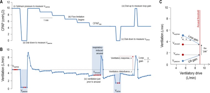
Study objectives: Obstructive sleep apnea (OSA) results from the interaction of several physiological traits; specifically a compromised upper airway anatomy and muscle function, and two key non-anatomical deficits: elevated loop gain and a low arousal threshold. Although continuous positive airway pressure (CPAP) is an efficacious treatment, it is often poorly tolerated. An alternative approach could involve administering therapies targeting the non-anatomic causes. However, therapies (oxygen or hypnotics) targeting these traits in isolation typically improve, but rarely resolve OSA. Therefore, our aim was to determine how the combination of oxygen and eszopiclone alters the phenotypic traits and OSA severity and to assess the baseline phenotypic characteristics of responders/nonresponders to combination therapy.

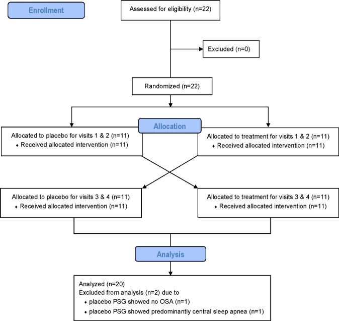
Methods: In a single-blinded randomized crossover study, 20 OSA patients received combination therapy (3 mg eszopiclone and 40% oxygen) versus placebo/sham air, with 1 w between conditions. Under each condition, we assessed the effects on OSA severity (clinical polysomnography) and the phenotypic traits causing OSA using CPAP manipulations (research polysomnography).

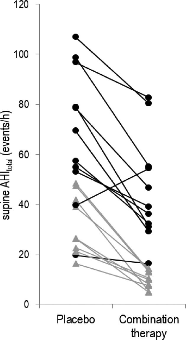
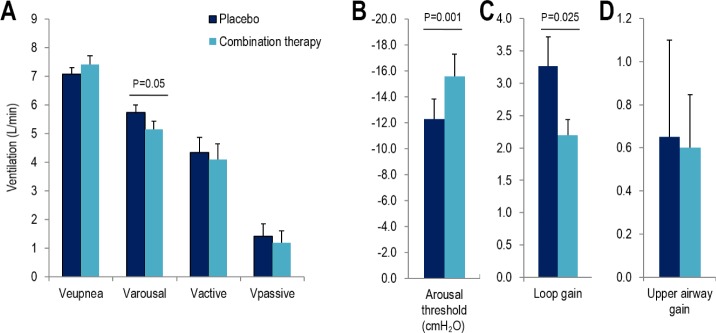
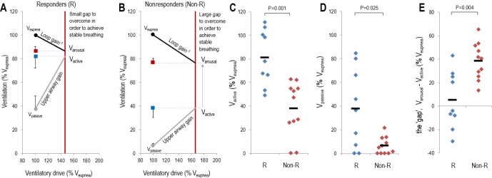
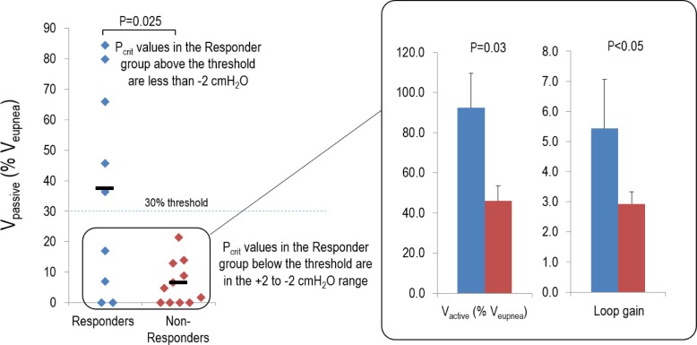
Results: Combination therapy reduced the apnea-hypopnea index (51.9 ± 6.2 vs. 29.5 ± 5.3 events/h; P < 0.001), lowered both the ventilation associated with arousal (5.7 ± 0.3 vs. 5.2 ± 0.3 L/min; P = 0.05) and loop gain (3.3 ± 0.5 vs. 2.2 ± 0.3; P = 0.025). Responders to therapy (apnea-hypopnea index reduced by > 50% to below 15 events/h; n = 9/20) had less severe OSA (P = 0.001), a less collapsible upper airway (P = 0.01) and greater upper airway muscle effectiveness (P = 0.002).

Conclusions: The combination of lowering loop gain and raising the arousal threshold is an effective therapy in patients whose anatomy is not severely compromised. Our work demonstrates that combining therapies that target multiple traits can resolve OSA in selected individuals.

Clinical trial registration: ClinicalTrials.gov, ID: NCT01633827.

Keywords: arousal threshold; loop gain; phenotyping; sleep apnea.


Figures

Figure 1
Figure 1
Flow diagram of the enrollment, randomization, and analysis procedures.
Figure 2
Figure 2
Technique for determining the physiological traits using continuous positive airway pressure (CPAP) dial-downs. The obstructive sleep apnea (OSA) traits are measured by manipulating CPAP during supine nonrapid eye movement (NREM) sleep (A) and measuring the changes in ventilation (B). After determining ventilation on optimum CPAP (Veupnea; i), the pressure was dialed down to measure passive V0 (ii), the ventilation at CPAP = 0 when pharyngeal muscles are inactive. CPAP was then lowered until flow limitation started (iii) and arousals occurred intermittently. Ventilation just prior to arousal (iv) was defined as the “ventilation causing arousal” (Varousal). During stable breathing between arousals, CPAP was dialed down or up to obtain active V0 (v) and loop gain (vi), respectively. Active V0 is the ventilation at CPAP = 0 when pharyngeal muscles are maximally activated. Loop gain is the ventilatory response (overshoot in ventilation above Veupnea) divided by the ventilatory disturbance (reduction in ventilation below Veupnea). (C) These four ventilations can be used to calculate the arousal threshold and upper airway response and illustrate how all four traits interact to manifest the absence or presence of OSA. After the patient's loop gain is known, the ventilation that causes arousal was translated into the ventilatory drive that causes the arousal, which is referred to as the arousal threshold (i.e., 9 L/min). When the arousal threshold is known, starting at the passive V0 we can say that if the ventilatory drive is increased to a level near the arousal threshold, then the pharyngeal muscles will activate and increase V0 to the measured level active V0. The slope of this line is called the upper airway gain (or response) and is a measure of the upper airway muscle function. The steeper this slope, the better the upper airway response to an increase in ventilatory drive, and the larger the distance between the active and passive V0. In order to avoid having OSA (i.e., achieve stable breathing), Vactive must be above Varousal.
Figure 3
Figure 3
Individual effects of combination therapy on obstructive sleep apnea (OSA) severity. Combination therapy significantly reduced the overall supine apnea-hypopnea index (AHI) in all but one individual. Notably this individual had a low loop gain at baseline and a robust upper airway muscle response. However, with therapy, for reasons that are unclear, combination therapy unexpectedly increased loop gain (by 150%) and attenuated their upper airway muscle function (by 84%), which consequently widened the gap between Varousal and Vactive: such a widening is consistent with the increase in OSA severity observed. Black circles represent those patients considered nonresponders to therapy, whereas gray triangles represent responders (see text for definitions of responders/nonresponders). Of note, in six patients (approximately one-third of all unselected patients) OSA was completely resolved (AHI < 10 events/h) with combination therapy.
Figure 4
Figure 4
The effect of combination therapy on the traits. Combination therapy significantly reduced Varousal (A) and loop gain (C) while also increasing the arousal threshold (B) (measured as nadir epiglottic pressure). Combination therapy did not alter any of the remaining traits: Veupnea, Vactive & Vpassive (A), upper airway gain (D).
Figure 5
Figure 5
Baseline physiological characteristics of responders to therapy. (A,B) The baseline group model diagrams for responders (R) and nonresponders (Non-R), respectively. Each model diagram illustrates how the four physiological traits interact to produce obstructive sleep apnea in each group. Data are expressed as a percentage of the eupneic ventilation (Veupnea). Responders to therapy had a higher maximum ventilation achievable without arousal (Vactive) (C) and a less collapsible airway under passive conditions (Vpassive) (D), which ultimately results in a smaller difference between Varousal and Vactive, called the physiologic “gap.” This small difference in responders means that only reductions in loop gain and increases in the arousal threshold are necessary for the intersection of the loop gain and upper airway gain lines to occur to the left of the arousal threshold line, thus yielding stable breathing.
Figure 6
Figure 6
Physiological characteristics of responders with a poor anatomy/collapsibility. Data are expressed as a percentage of the eupneic ventilation (Veupnea). Individuals with a Vpassive below 30% of their eupneic ventilation (Veupnea) were classified as having a poor anatomy. Note that four of nine responders fit this category, whereas all of the nonresponders had a poor anatomy. When comparing the physiological differences between the responders (shown in blue) and nonresponders (shown in red) in this subset (see inset), responders had a higher maximum ventilation achievable without arousal (Vactive) and a higher loop gain.

References

    1. Somers VK, Dyken ME, Clary MP, Abboud FM. Sympathetic neural mechanisms in obstructive sleep apnea. J Clin Invest. 1995;96:1897–904. - PMC - PubMed
    1. Nieto F, Young TB, Lind BK, et al. Association of sleep-disordered breathing, sleep apnea, and hypertension in a large community-based study. Sleep Heart Health Study. JAMA. 2000;283:1829–36. - PubMed
    1. Brooks D, Horner RL, Kozar LF, Render-Teixeira CL, Phillipson EA. Obstructive sleep apnea as a cause of systemic hypertension. Evidence from a canine model [see comments] J Clin Invest. 1997;99:106–9. - PMC - PubMed
    1. Peppard P, Young T, Palta M, Skatrud J. Prospective study of the association between sleep disordered breathing and hypertension. N Engl J Med. 2000;342:1378–84. - PubMed
    1. Hung J, Whitford EG, Parsons RW, Hillman DR. Association of sleep apnoea with myocardial infarction in men [see comments] Lancet. 1990;336:261–4. - PubMed

Publication types

MeSH terms

Associated data