

Superficial temporal artery pseudoaneurysm: what is the role of ultrasound?
- PMID: 27635165
- PMCID: PMC5005213
- DOI: 10.1007/s40477-016-0211-8
Superficial temporal artery pseudoaneurysm: what is the role of ultrasound?
Abstract
Purpose: The superficial temporal artery (STA) is one of the terminal branches of the external carotid artery; STA pseudoaneurysms are uncommon vascular lesion, generally subsequent to blunt or penetrating trauma that could represent a trick for radiologist, especially when the only anamnestic information is "palpable superficial swelling". In this article, we describe our ultrasonographic experience about STA pseudoaneurysm reporting several cases with different etiopatogenesis.
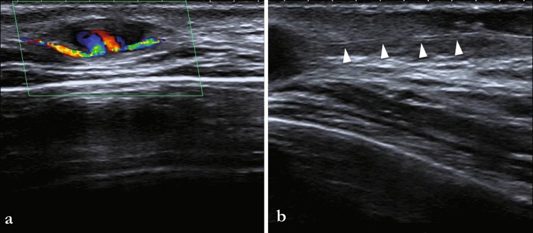
Methods: Between January 2004 and March 2015 six patients (4F and 2M; aged 15-55 years, mean 36 year) presented at our department with superficial palpable swelling in temporal region (four with trauma history, two with iatrogenic cause) underwent to ultrasonographic study to assess the presence of STA pseudoaneurysm. Ultrasonographic findings suggestive of pseudoaneurysm was a well-defined, pulsatile, anechoic mass in B-mode, a swirling or disorganized pattern of blood flow in the lesion with demonstration of direct communication between arterial lumen and pseudoaneurysm at colour-Doppler and a typical to-and-fro waveform on pseudoaneurysm neck at pulsed-Doppler.
Results: B-mode proves the presence of anechoic mass in five on six patients. Colour-Doppler demonstrates the presence of flow inside the lesion in five patients and a direct communication in all patients. To-and-fro typical waveform has been demonstrated in five patients. Ultrasound made diagnosis in all patients with a sensibility and specificity of 100 %.
Conclusion: US is the imaging modality of choice, since it can provide detailed information about vascular anatomy without incurring the risks of invasive methods like angiography or radiation.
Scopo: L’arteria temporale superficiale (STA) è uno dei rami terminali della carotide esterna. Gli pseudoaneurismi dell’STA sono poco frequenti, generalmente successivi a traumi chiusi o penetranti, potendo trarre in inganno il radiologo soprattutto quando l’unica informazione anamnestica è “rigonfiamento superficiale palpabile”. In quest’articolo descriviamo la nostra esperienza ultrasonografica riguardo gli pseudoaneurismi dell’STA riportando diversi casi ad eziopatogenesi diversa.
Metodi: Tra il Gennaio 2004 e Marzo 2015 6 pazienti (4F E 2M; Età 15–55 anni, media 36) giunti al nostro dipartimento per un rigonfiamento superficiale palpabile in regione temporale (4 con storia di trauma, 2 per cause iatrogene) vengono sottoposti ad esame ecografico per valutare la presenza di pseudoaneurismi dell’STA. I reperti ultrasonografici suggestivi per pseudoaneurisma sono stati: una massa ben definita, anecogena e pulsatile in B-mode, un pattern di flusso turbolento all’interno della lesione con la dimostrazione di una diretta comunicazione tra il lume arterioso e lo pseudoaneurisma al color-Doppler e un tipico spettro flussimetrico “to-and-fro” in corrispondenza del collo aneurismatico.
Risultati: B-mode ha mostrato la presenza di una massa anecogena in 5 su 6 pazienti. Il color-Doppler ha dimostrato la presenza di flusso all’interno della lesione in 5 pazienti e una diretta comunicazione in tutti i pazienti. Lo spettro “to-and-fro” è stato dimostrato in 5 pazienti. L’ecografia ha permesso la diagnosi in tutti i pazienti con una sensibilità e specificità pari al 100 %.
Conclusioni: L’ecografia rappresenta la modalità di imaging di scelta, fornendo informazioni dettagliate sull’anatomia vascolare senza incorrere nei rischi di metodiche invasive come l’angiografia o all’utilizzo di radiazioni.
Keywords: Colour Doppler; Pseudoaneurysm; Sonography; Superficial temporal artery.
Conflict of interest statement
Compliance with ethical standards Human and animal rights All ethical procedures performed in studies involving human participants were in accordance with the ethical standards of the institutional and/or national research committee and with the 1964 Helsinki declaration and its later amendments or comparable ethical standards. Informed consent All patients provided written informed consent to enrolment in the study and to the inclusion in this article of information that could potentially lead to their identification. Conflict of interest The authors declare that they have no conflict of interest.
Figures


References
-
- Bartholin T. Epistolarium medicinalum centuria. The Hague: Comitum; 1644. p. 53.
MeSH terms
LinkOut - more resources
Full Text Sources
Other Literature Sources
