. 2016 Sep 15;63(6):1034-43.
doi: 10.1016/j.molcel.2016.08.013.

Coincident Phosphatidic Acid Interaction Restrains Drp1 in Mitochondrial Division

Affiliations

Coincident Phosphatidic Acid Interaction Restrains Drp1 in Mitochondrial Division

Yoshihiro Adachi et al. Mol Cell. .

Abstract

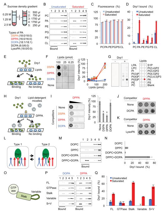
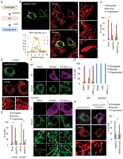
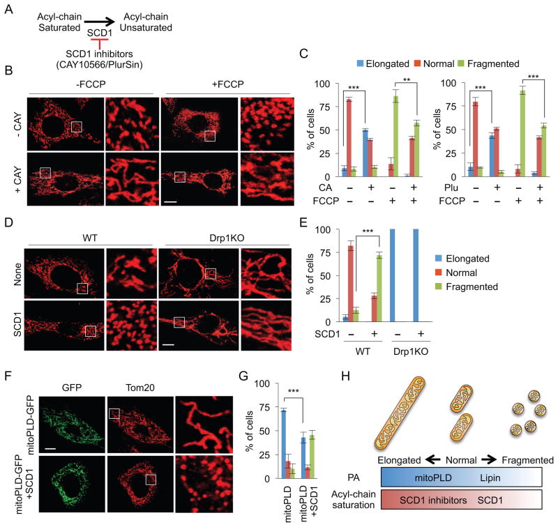
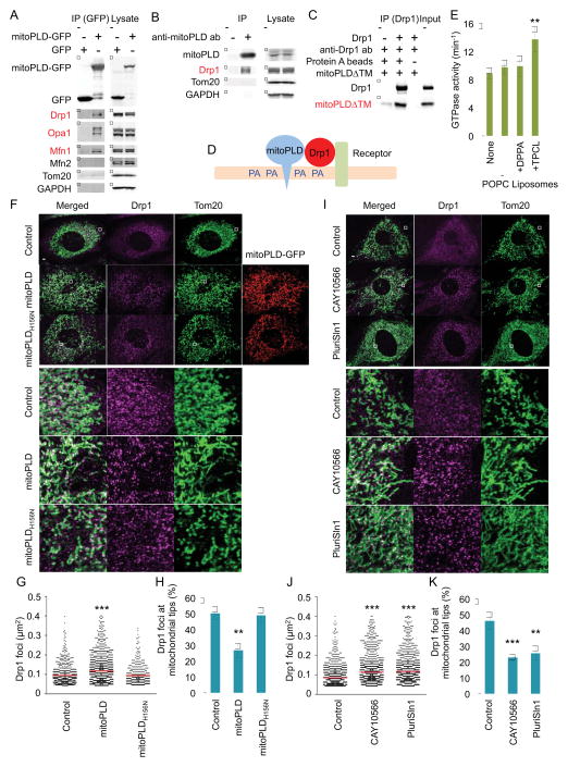
Mitochondria divide to control their size, distribution, turnover, and function. Dynamin-related protein 1 (Drp1) is a critical mechanochemical GTPase that drives constriction during mitochondrial division. It is generally believed that mitochondrial division is regulated during recruitment of Drp1 to mitochondria and its oligomerization into a division apparatus. Here, we report an unforeseen mechanism that regulates mitochondrial division by coincident interactions of Drp1 with the head group and acyl chains of phospholipids. Drp1 recognizes the head group of phosphatidic acid (PA) and two saturated acyl chains of another phospholipid by penetrating into the hydrophobic core of the membrane. The dual phospholipid interactions restrain Drp1 via inhibition of oligomerization-stimulated GTP hydrolysis that promotes membrane constriction. Moreover, a PA-producing phospholipase, MitoPLD, binds Drp1, creating a PA-rich microenvironment in the vicinity of a division apparatus. Thus, PA controls the activation of Drp1 after the formation of the division apparatus.


Figures

Figure 1
Figure 1. Coincident interaction of Drp1 with saturated PA
(A) Liposome floatation assays. After ultracentrifugation, we collected five fractions from the top and then analyzed the Drp1-liposome association in (B). Rhodamine-PE fluorescence in (C) and Drp1 band intensity in (D) were normalized to the volume of fractions (Mean ± SEM; n = 3). (E) Lipid dot-blot assays. (F and G) His6-Drp1 was incubated with PVDF membranes spotted for the indicated lipids in (F) or PIP strip (Echelon P-6001) in (G), and then detected Drp1-lipid interactions using anti-Drp1 antibodies (Mean ± SEM; n = 3). (H) Lipid competition assays. (I–K) Drp1 was incubated with membranes spotted for DPPA in the presence of the indicated lipids. (L) Model for Drp1-phospholipid interactions. (M and N) We analyzed interactions of His6-Drp1 with the indicated liposomes using floatation assays. (O) Domain structure of Drp1. (P and Q) Different domains were analyzed in the liposome floatation assay (Mean ± SEM; n = 3). “n” indicates the number of independent experiments. Student’s t-test: *p< 0.05. See also Figure S1.
Figure 2
Figure 2. Suppression of Drp1-mediated mitochondrial division by PA
(A) Conversion of lipids mediated by MitoPLD and lipin. MEFs were transfected with MitoPLD-GFP in (B) or catalytically inactive MitoPLDH156N-GFP in Supplementary Figure 3. We quantified GFP intensity along the lines #1–3. Mitochondrial shape was visualized using immunofluorescence with anti-Tom20 antibodies. The boxed regions are enlargements. (C) Quantification of mitochondrial morphology in cells modestly expressing MitoPLD-GFP or MitoPLDH156N-GFP. Mean ± SD (n = 3, 90 cells). (D and E) Mitochondria were analyzed in MEFs modestly expressing MitoPLD-GFP or MitoPLDH156N-GFP after FCCP treatment for 30 min. The boxed regions are enlargements. Mean ± SD (n = 3, 90 cells). (F) WT and Drp1KO MEFs were transfected with HA-lipin1b, which converts PA to diacylglycerol, or enzymatically inactive HA-lipinD712A. The cells were subjected to immunofluorescence microscopy with anti-Tom20 antibodies. (G) We quantified mitochondrial shape. Mean ± SEM (n = 3, 90 cells). (H and I) Mitochondrial morphology was examined in MEFs modestly expressing MitoPLD-GFP in the presence or absence of HA-lipin using immunofluorescence microscopy. Mean ± SD (n = 3, 90 cells). “n” indicates the number of independent experiments. Student’s t-test: ***p< 0.001. Bar, 10 μm. See also Figure S2, S3 and S4.
Figure 3
Figure 3. Reduction of mitochondrial division via saturation of acyl chains
(A) SCD1 mediates the desaturation of fatty acid acyl chains. (B and C) After incubation with SCD1 inhibitors, CAY10566 (CAY) and PluriSln1 (Plu), WT MEFs were treated with 10 μM FCCP for 30 min. Mitochondrial shape was analyzed using immunofluorescence microscopy with anti-Tom20 antibodies. Mean ± SD (n = 3, 90 cells). (D and E) WT and Drp1KO MEFs were transfected with SCD1 and analyzed for mitochondrial morphology using immunofluorescence microscopy. Mean ± SD (n = 3, 90 cells). (F and G) We examined mitochondria in WT MEFs mildly expressing MitoPLD-GFP in the presence or absence of SCD1. Mean ± SD (n = 3, 90 cells). (H) Summary of the effects of PA and saturated acyl chain on mitochondrial shape. “n” indicates the number of independent experiments. Student’s t-test: **p< 0.01; ***p< 0.001. Bar, 10 μm.
Figure 4
Figure 4. Mechanism of phospholipid-mediated suppression of mitochondrial division
(A) Cells expressing MitoPLD-GFP or GFP were subjected to immunoprecipitation using anti-GFP antibodies. Drp1, Mfn1 and Opa1 specifically bind mitoPLD-GFP. (B) Mouse testes were subjected to immunoprecipitation using anti-MitoPLD antibodies. Drp1 specifically bind MitoPLD. (C) A FLAG-tagged cytoplasmic domain of MitoPLD (mitoPLDΔTM) was incubated with His6-Drp1, and His6-Drp1 was pulled down with anti-Drp1 antibodies. Drp1 directly binds mitoPLDΔTM. (D) Model for inhibitory lipid microenvironment created by the Drp1-MitoPLD interaction. (E) The GTPase activity of His6-Drp1 was measured in the presence of the indicated liposomes. Saturated PA containing liposomes does not stimulate the GTPase activity of Drp1. Mean ± SEM (n = 5). (F–K), WT MEFs expressing MitoPLD-GFP or MitoPLDH156N-GFP (F, G and H) or treated with SCD1 inhibitors (I, J and K) were subjected to immunofluorescence microscopy with antibodies specific for Tom20 and Drp1. The sizes of the Drp1 foci in the mitochondria were quantified (G and J). Red lines correspond to means (more than 300 Drp1 foci were analyzed). The relative abundance of Drp1 foci associated with the tips of mitochondrial tubules was determined (H and K). Mean ± SEM (n = 10 cells). Student’s t-test: **p< 0.01, ***p< 0.01. Bar, 10 μm.

Comment in

References

    1. Baba T, Kashiwagi Y, Arimitsu N, Kogure T, Edo A, Maruyama T, Nakao K, Nakanishi H, Kinoshita M, Frohman MA, et al. Phosphatidic acid (PA)-preferring phospholipase A1 regulates mitochondrial dynamics. The Journal of biological chemistry. 2014;289:11497–11511. - PMC - PubMed
    1. Ben-David U, Gan QF, Golan-Lev T, Arora P, Yanuka O, Oren YS, Leikin-Frenkel A, Graf M, Garippa R, Boehringer M, et al. Selective elimination of human pluripotent stem cells by an oleate synthesis inhibitor discovered in a high-throughput screen. Cell Stem Cell. 2013;12:167–179. - PubMed
    1. Bleazard W, McCaffery JM, King EJ, Bale S, Mozdy A, Tieu Q, Nunnari J, Shaw JM. The dynamin-related GTPase Dnm1 regulates mitochondrial fission in yeast. Nat Cell Biol. 1999;1:298–304. - PMC - PubMed
    1. Bui HT, Shaw JM. Dynamin assembly strategies and adaptor proteins in mitochondrial fission. Current biology : CB. 2013;23:R891–899. - PMC - PubMed
    1. Bustillo-Zabalbeitia I, Montessuit S, Raemy E, Basanez G, Terrones O, Martinou JC. Specific interaction with cardiolipin triggers functional activation of Dynamin-Related Protein 1. PLoS One. 2014;9:e102738. - PMC - PubMed

Publication types

MeSH terms