. 2016 Sep 16;11(9):e0162476.
doi: 10.1371/journal.pone.0162476. eCollection 2016.

Relationship between Human Pupillary Light Reflex and Circadian System Status

Affiliations

Relationship between Human Pupillary Light Reflex and Circadian System Status

Maria Angeles Bonmati-Carrion et al. PLoS One. .

Abstract

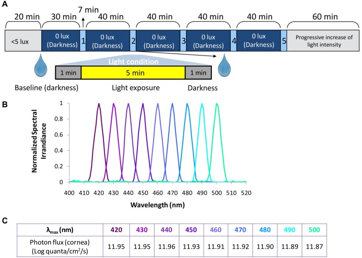
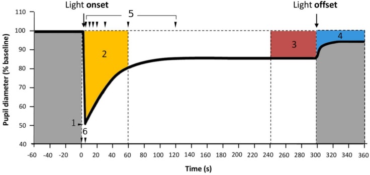
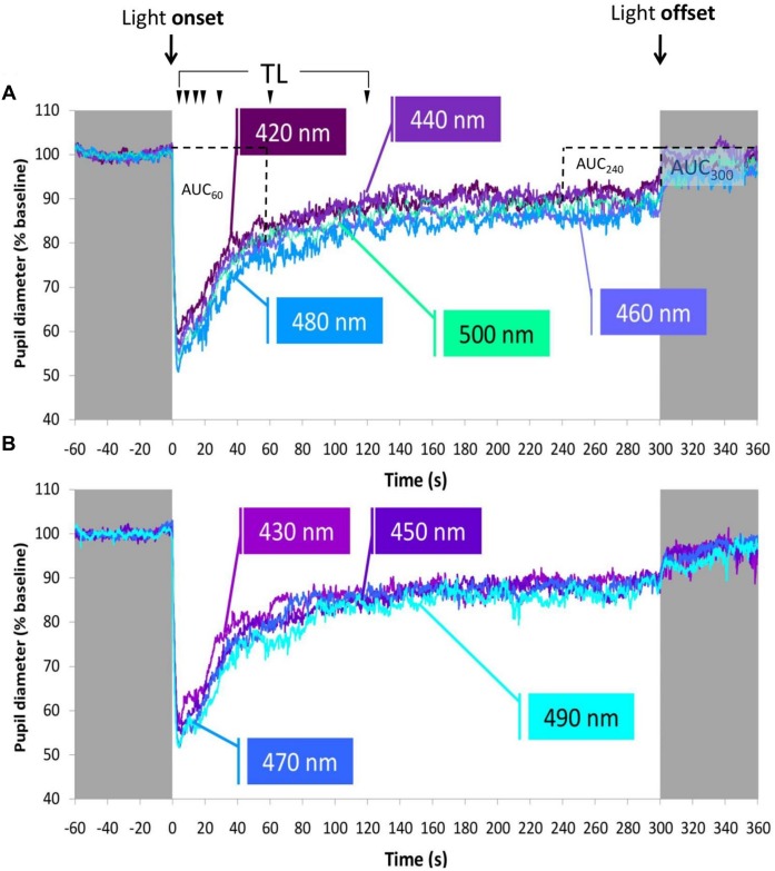
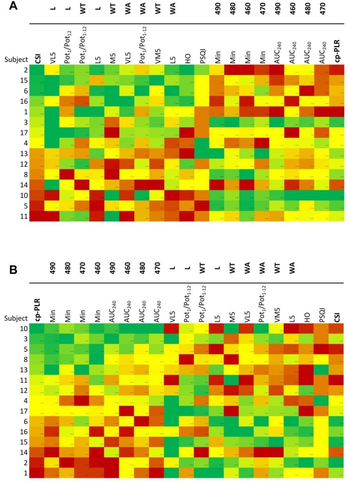
Intrinsically photosensitive retinal ganglion cells (ipRGCs), whose photopigment melanopsin has a peak of sensitivity in the short wavelength range of the spectrum, constitute a common light input pathway to the olivary pretectal nucleus (OPN), the pupillary light reflex (PLR) regulatory centre, and to the suprachiasmatic nuclei (SCN), the major pacemaker of the circadian system. Thus, evaluating PLR under short wavelength light (λmax ≤ 500 nm) and creating an integrated PLR parameter, as a possible tool to indirectly assess the status of the circadian system, becomes of interest. Nine monochromatic, photon-matched light stimuli (300 s), in 10 nm increments from λmax 420 to 500 nm were administered to 15 healthy young participants (8 females), analyzing: i) the PLR; ii) wrist temperature (WT) and motor activity rhythms (WA), iii) light exposure (L) pattern and iv) diurnal preference (Horne-Östberg), sleep quality (Pittsburgh) and daytime sleepiness (Epworth). Linear correlations between the different PLR parameters and circadian status index obtained from WT, WA and L recordings and scores from questionnaires were calculated. In summary, we found markers of robust circadian rhythms, namely high stability, reduced fragmentation, high amplitude, phase advance and low internal desynchronization, were correlated with a reduced PLR to 460-490 nm wavelengths. Integrated circadian (CSI) and PLR (cp-PLR) parameters are proposed, that also showed an inverse correlation. These results demonstrate, for the first time, the existence of a close relationship between the circadian system robustness and the pupillary reflex response, two non-visual functions primarily under melanopsin-ipRGC input.


Conflict of interest statement

The authors have declared that no competing interests exist.

Figures

Fig 1
Fig 1. In-laboratory protocol.
(A) Light conditions tested including the study protocol, (B) normalized spectra and (C) their corresponding photon fluxes at the cornea. Drop symbol shows the time when the pupil dilator, tropicamide, was applied.
Fig 2
Fig 2. Parameters assessed for PLR.
1: Minimum diameter, expressed as percentage constriction; 2: Area Under the Curve from 0 to 60 seconds of light exposure (AUC60, orange); 3: Area Under the Curve from 240 to 300 seconds of light exposure (AUC240, in red); 4: Area Under the Curve from light offset to the end of the recording (from 300 to 360 seconds of recording) (AUC300, blue); 5: Percentage pupil constriction. Each arrow indicates, from left to right, TL5, TL10, TL15, TL20, TL30, TL60 and TL120, respectively, thus the percentage of pupil constriction at 5, 10, 15, 20, 30, 60 and 120 seconds after light onset.; 6: Time from light onset to the minimum pupil diameter reached during pupil constriction.
Fig 3
Fig 3. Ambulatory circadian monitoring.
Left panel: nine-day averaged recording for (A) wrist temperature, (B) wrist acceleration, and (C) light exposure, from 15 subjects. Right panel: averaged mean waveforms (n = 15) for (D) wrist temperature, (E) wrist acceleration, and (F) light exposure. Data are expressed as mean ± SEM.
Fig 4
Fig 4. Averaged pupil recordings under the different lights tested (n = 15).
Averaged pupil recordings under (A) 420, 440, 460, 480, 500 nm and (B) 430, 450, 470 and 490 nm lights. Grey areas represent darkness. On the top, light onset and offset are indicated. Metrics (see Fig 2) are also indicated here. TL: Each arrow indicates, from left to right, TL5, TL10, TL15, TL20, TL30, TL60 and TL120, respectively, and the percent baseline pupil diameter at 5, 10, 15, 20, 30, 60 and 120 seconds after light onset. AUC60 (Area Under the Curve from 0 to 60 seconds of light exposure), AUC240 (Area Under the Curve from 240 to 300 seconds of light exposure) and AUC300 (Area Under the Curve from 300 to 360 seconds of recording, after light offset in darkness) are indicated in Fig 4A.
Fig 5
Fig 5. Macroarrays according to high and low cp-PLR and CSI parameters.
Graphic matrix for subjects (n = 15) sorted by (A) CSI (circadian status index) and (B) by cp-PLR (circadian photoreception PLR). Colour scale corresponds to the relative magnitude for each variable and participant: reddish colours indicate lower scores for both CSI (worse circadian system status) and PLR parameters (less constriction) with those wavelengths in the circadian range (460-490nm). For details on the cp-PLR and CSI indexes calculation see Materials and Methods. The variables included in the macroarrays are wrist temperature (WT), wrist acceleration (WA) and light exposure (L) from the circadian analysis and normalized minimum pupil diameter expressed as pupil constriction and AUC240. M5, the mid-point time of the five consecutive hours with the highest values; VM5, value for M5; L5, the mid-point time of the five consecutive hours with the lowest values; VL5, value for L5; HO, Horne-Östberg Morningness-Eveningness Questionnaire score; PSQI, Pittsburgh Sleep Quality Index; Min, minimum pupil diameter (expressed as pupil constriction); AUC240, Area Under the Curve during the last minute (240 seconds) of light exposure. See Materials and Methods for the calculation details.

References

    1. Provencio I, Rodriguez IR, Jiang G, Hayes WP, Moreira EF, Rollag MD. A novel human opsin in the inner retina. J Neurosci. 2000;20: 600–5. - PMC - PubMed
    1. Berson DM, Dunn FA, Takao M. Phototransduction by retinal ganglion cells that set the circadian clock. Science. 2002;295: 1070–3. - PubMed
    1. Hattar S, Liao HW, Takao M, Berson DM, Yau KW. Melanopsin-containing retinal ganglion cells: architecture, projections, and intrinsic photosensitivity. Science. 2002;295: 1065–70. - PMC - PubMed
    1. Panda S, Sato TK, Castrucci AM, Rollag MD, DeGrip WJ, Hogenesch JB, et al. Melanopsin (Opn4) requirement for normal light-induced circadian phase shifting. Science. 2002;298: 2213–6. - PubMed
    1. Lucas RJ, Hattar S, Takao M, Berson DM, Foster RG, Yau K-W. Diminished pupillary light reflex at high irradiances in melanopsin-knockout mice. Science. 2003;299: 245–7. - PubMed