. 2016 Dec;25(12):2290-2296.
doi: 10.1002/pro.3042. Epub 2016 Sep 26.

Solution structure of protein synthesis initiation factor 1 from Pseudomonas aeruginosa

Affiliations

Solution structure of protein synthesis initiation factor 1 from Pseudomonas aeruginosa

Yanmei Hu et al. Protein Sci. 2016 Dec.

Abstract

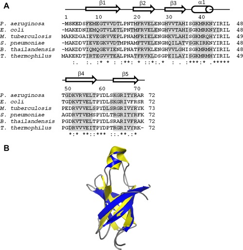
Pseudomonas aeruginosa is an opportunistic bacterial pathogen and a primary cause of nosocomial infection in humans. The rate of antibiotic resistance in P. aeruginosa is increasing worldwide leading to an unmet need for discovery of new chemical compounds distinctly different from present antimicrobials. Protein synthesis is an essential metabolic process and a validated target for the development of new antibiotics. Initiation factor 1 from P. aeruginosa (Pa-IF1) is the smallest of the three initiation factors that act to establish the 30S initiation complex during initiation of protein biosynthesis. Here we report the characterization and solution NMR structure of Pa-IF1. Pa-IF1 consists of a five-stranded β-sheet with an unusual extended β-strand at the C-terminus and one short α-helix arranged in the sequential order β1-β2-β3-α1-β4-β5. The structure adopts a typical β-barrel fold and contains an oligomer-binding motif. A cluster of basic residues (K39, R41, K42, K64, R66, R70, and R72) located on the surface of strands β4 and β5 near the short α-helix may compose the binding interface with the 30S subunit.

Keywords: NMR structure; Pseudomonas aeruginosa; translation initiation factor 1; β-barrel motif.


Figures

Figure 1
Figure 1
(A) Alignment (Clustal Omega) of the primary sequence of Pa‐IF1 with those of other bacterial homologs from: E. coli (PDB 1AH9), M. tuberculosis (PDB 3I4O), S. pneumoniae (PDB 4QL5), B. thailandensis (PDB 2N3S), T. thermophilus (PDB 1HR0). Secondary structural elements, highlighted in gray, were derived from the PDB structures. The secondary structure elements of Pa‐IF1 are indicated schematically above the sequence. (B) Overlay of Pa‐IF1 (secondary structures in blue) and IF1 from E. coli (PDB 1AH9, secondary structures in yellow).
Figure 2
Figure 2
Characterization of P. aeruginosa IF1. (A) P. aeruginosa IF1 functions alone in the P. aeruginosa A/T assay to prevent formation of 70s ribosomes and, therefore, decreases poly‐Phe synthesis; (B) IF1 enhances the inhibition of the formation of 70S ribosomes in the presence of 10 μM P. aeruginosa IF3.
Figure 3
Figure 3
The NMR longitudinal relaxation (T 1) and transverse relaxation (T 2) of the residue‐specific backbone amide 15N of Pa‐IF1 at 298 K. The secondary structure elements are indicated schematically at the top of the figure.
Figure 4
Figure 4
NMR‐derived Structures of Pa‐IF1. (A) Superposition of main chain atoms of 15‐lowest‐energy conformers with an RMSD of 0.3 Å (main chain atoms). The first six N‐terminal residues are not folded in the structure. (B) Ribbon representation of the energy‐minimized average main chain structure with secondary structures labeled (α‐helix: orange; β strands: blue). (C) Surface presentation of the electrostatic potential (±1 kT/e) generated using the Adaptive Poisson‐Boltzmann Equation (APBS) plugin in PyMOL with ionic strength 0.15 M and views at 90° and 180° rotation around the longitudinal axis. Red and blue colored regions denote negative and positive charges, respectively.

References

    1. Stover CK, Pham XQ, Erwin AL, Mizoguchi SD, Warrener P, Hickey MJ, Brinkman FS, Hufnagle WO, Kowalik DJ, Lagrou M, Garber RL, Goltry L, Tolentino E, Westbrock‐Wadman S, Yuan Y, Brody LL, Coulter SN, Folger KR, Kas A, Larbig K, Lim R, Smith K, Spencer D, Wong GK, Wu Z, Paulsen IT, Reizer J, Saier MH, Hancock RE, Lory S, Olson MV (2000) Complete genome sequence of Pseudomonas aeruginosa PAO1, an opportunistic pathogen. Nature 406:959–964. - PubMed
    1. Hirsch EB, Tam VH (2010) Impact of multidrug‐resistant Pseudomonas aeruginosa infection on patient outcomes. Expert Rev Pharmacoecon Outcomes Res 10:441–451. - PMC - PubMed
    1. McCoy LS, Xie Y, Tor Y (2011) Antibiotics that target protein synthesis. RNA 2:209–232. - PubMed
    1. Laursen BS, Sorensen HP, Mortensen KK, Sperling‐Petersen HU (2005) Initiation of protein synthesis in bacteria. Microbiol Mol Biol Rev 69:101–123. - PMC - PubMed
    1. Julian P, Milon P, Agirrezabala X, Lasso G, Gil D, Rodnina MV, Valle M (2011) The cryo‐EM structure of a complete 30S translation initiation complex from Escherichia coli . PLoS Biol 9:e1001095. - PMC - PubMed

MeSH terms

Substances

LinkOut - more resources