. 2016 Oct 18;7(42):67934-67947.
doi: 10.18632/oncotarget.11982.

Chromatin determinants of the inner-centromere rely on replication factors with functions that impart cohesion

Affiliations

Chromatin determinants of the inner-centromere rely on replication factors with functions that impart cohesion

Takuya Abe et al. Oncotarget. .

Abstract

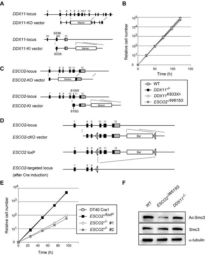
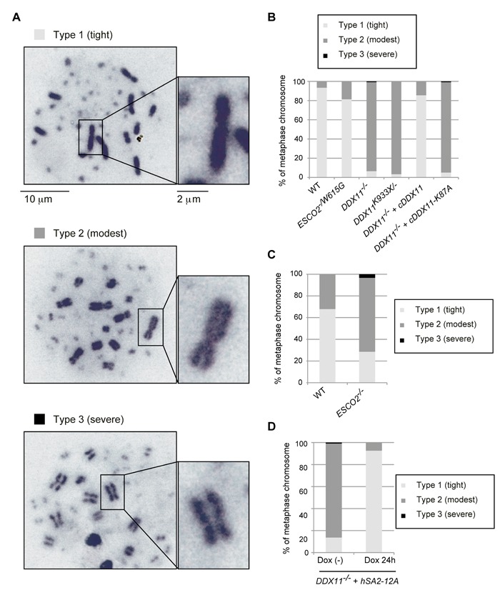
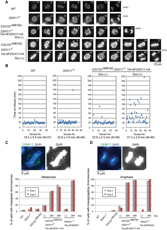
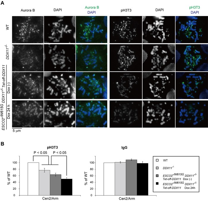
Replication fork-associated factors promote genome integrity and protect against cancer. Mutations in the DDX11 helicase and the ESCO2 acetyltransferase also cause related developmental disorders classified as cohesinopathies. Here we generated vertebrate model cell lines of these disorders and cohesinopathies-related genes. We found that vertebrate DDX11 and Tim-Tipin are individually needed to compensate for ESCO2 loss in chromosome segregation, with DDX11 also playing complementary roles with ESCO2 in centromeric cohesion. Our study reveals that overt centromeric cohesion loss does not necessarily precede chromosome missegregation, while both these problems correlate with, and possibly originate from, inner-centromere defects involving reduced phosphorylation of histone H3T3 (pH3T3) in the region. Interestingly, the mitotic pH3T3 mark was defective in all analyzed replication-related mutants with functions in cohesion. The results pinpoint mitotic pH3T3 as a postreplicative chromatin mark that is sensitive to replication stress and conducts with different kinetics to robust centromeric cohesion and correct chromosome segregation.

Keywords: DDX11; Tim-Tipin; inner-centromere; replication stress; sister chromatid cohesion; Chromosome Section.


Conflict of interest statement

CONFLICTS OF INTEREST

The authors declare that they have no conflict of interest.

Figures

Figure 1
Figure 1. Establishment and general characterization of WABS- and RBS-model DT40 lines
A. Schematic representation of gene-targeting KO and KI constructs for DDX11. Black-filled rectangles indicate exons, and “Marker” indicates drug resistance genes. B., E. Growth curves. C.-D. Schematic representation of KO and KI constructs (C) and conditional gene-targeting constructs (D) for ESCO2. Black-filled rectangles and Bsr indicate exons and the Blasticidin S-resistance gene, respectively. F. Western blotting from total cell lysates for the markers indicated. The results were confirmed with lysates from an independent biological experiment.
Figure 2
Figure 2. Cohesion defects in various DDX11 and ESCO2 mutants
A.-D. Chromosomes from metaphase spreads were classified in three groups (A), and more than 100 metaphase cells were analyzed for each genotype (B-D). Independently prepared slides, from a different biological experiment, were used to confirm the trend. (D) DDX11 cells with hSA2-12A were incubated with or without Dox, to induce expression of hSA2-12A, for 24 h and metaphase spread samples were examined as in (B-C).
Figure 3
Figure 3. Synthetic lethality between DDX11−/− and ESCO2−/W615G mutations
A. Depletion of DDX11-HA protein and measurement of Ac-Smc3, Smc3, and α-tubulin (loading control) by Western blotting. The results were confirmed with lysates from an independent biological experiment. B. Growth curves. Dox was added at time 0. C.-D. Metaphase spreads examined as in Figure 2B with more than 100 metaphases examined for each genotype and independently prepared slides, from a different biological experiment, used to confirm the trend. E. Metaphase spread samples were prepared by the cytospin method after incubation with 0.1 μg/ml of colcemid for 1 h, and metaphase spread samples were prepared by the cytospin method. The distances between CENP-T signals were measured for more than 275 chromosomes. The same trend was confirmed from an independent biological experiment. p values were calculated by Student's t-test.
Figure 4
Figure 4. ESCO2−/W615G DDX11−/− cells show mitotic delays and chromosome missegregation
A. Dynamics of mitotic chromosomes in the indicated cell lines. Live cell imaging was initiated 24 h after Dox addition and continued until 32 h. B. Quantification of the time for progression from prophase to telophase. N represents the number of cells examined, the average time required to complete mitosis is indicated under the panels. C.-D. Misaligned chromosomes in metaphase (C) or missegregating chromosomes in anaphase (D). At least 100 cells for the metaphase plot and 50 cells for the anaphase plot were analyzed for each experiment. The results of two independent experiments are plotted.
Figure 5
Figure 5. ESCO2−/W615G DDX11−/− cells show inner centromere structural defects
A. Localization of Aurora B and phosphorylated-Histone H3T3 (pH3T3) from samples prepared as in Figure 3E. B. ChIP-qPCR of pH3T3 at a centromeric (CEN) region. The ratios of pH3T3 level between Cen2 and MHM repeats (Arm) were normalized by those of Input samples. Simultaneously immunoprecipitated DNAs with control IgG antibody and anti-pH3T3-coupled beads were amplified, and the ratios were calculated in the same way. Specific amplification of the centromeric regions and MHM repeats was controlled by PCR. Experiments were repeated three times in independent biological experiments. Bars indicate standard deviations. p values were calculated by Student's t-test.
Figure 6
Figure 6. Tim-Tipin pathway is essential in ESCO2−/W615G cells
A. Growth curves. B. Chromosomes were examined in the indicated genotypes as in Fig. 4D. 50 cells for each experiment were analyzed. The results of two independent experiments are plotted. C. Metaphase spreads, from cells incubated with Dox for 72 h, were classified for cohesion defects as in Figure 2B. More than 100 metaphase cells were examined. D. Localization of pH3T3 as in Figure 5A.

References

    1. Branzei D, Foiani M. Maintaining genome stability at the replication fork. Nat Rev Mol Cell Biol. 2010;11:208–219. - PubMed
    1. Moldovan GL, Pfander B, Jentsch S. PCNA, the maestro of the replication fork. Cell. 2007;129:665–679. - PubMed
    1. Leman AR, Noguchi E. Local and global functions of Timeless and Tipin in replication fork protection. Cell Cycle. 2012;11:3945–3955. - PMC - PubMed
    1. Bharti SK, Khan I, Banerjee T, Sommers JA, Wu Y, Brosh RM., Jr Molecular functions and cellular roles of the ChlR1 (DDX11) helicase defective in the rare cohesinopathy Warsaw breakage syndrome. Cell Mol Life Sci. 2014;71:2625–2639. - PMC - PubMed
    1. Cali F, Bharti SK, Di Perna R, Brosh RM, Jr, Pisani FM. Tim/Timeless, a member of the replication fork protection complex, operates with the Warsaw breakage syndrome DNA helicase DDX11 in the same fork recovery pathway. Nucleic Acids Res. 2015;44:705–717. - PMC - PubMed

MeSH terms

Supplementary concepts

LinkOut - more resources