. 2016 Nov 8;7(45):72807-72818.
doi: 10.18632/oncotarget.12020.

SASH1 mediates sensitivity of breast cancer cells to chloropyramine and is associated with prognosis in breast cancer

Affiliations

SASH1 mediates sensitivity of breast cancer cells to chloropyramine and is associated with prognosis in breast cancer

Joshua T Burgess et al. Oncotarget. .

Abstract

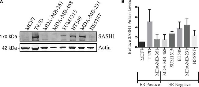
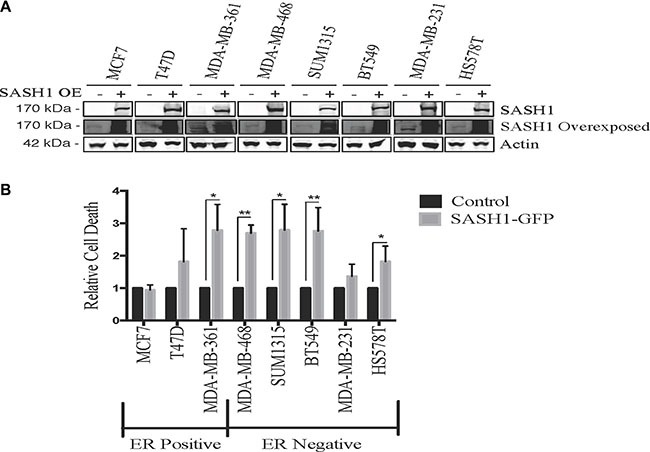
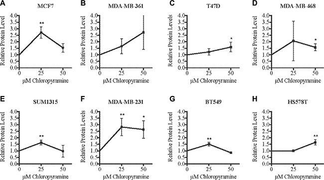
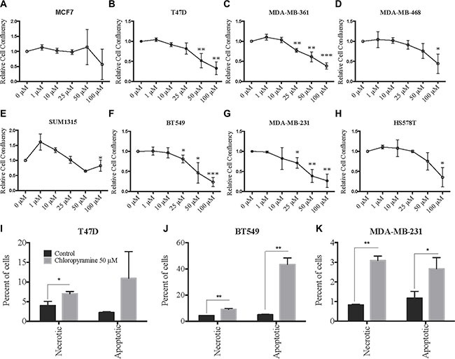
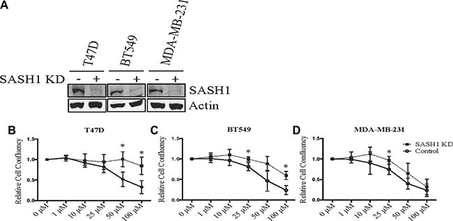
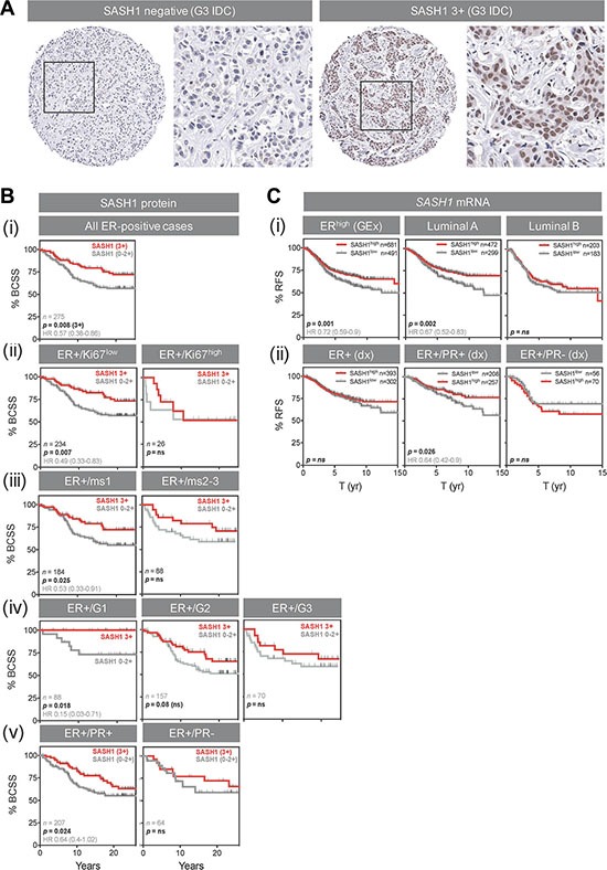
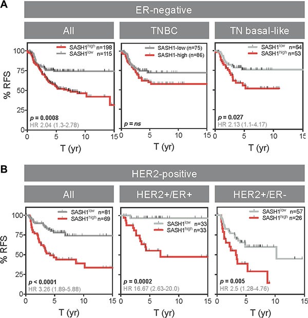
Expression of the SASH1 protein is reduced in a range of human cancers and has been implicated in apoptotic cancer cell death. This study investigated whether increasing SASH1 expression could be a useful therapeutic strategy in breast cancer. Ectopic SASH1 expression increased apoptosis in 7/8 breast cancer cell lines. Subsequent in silico connectivity screening demonstrated that the clinically approved antihistamine drug, chloropyramine, increased SASH1 mRNA levels. Chloropyramine has previously been shown to have anti-tumour activity in breast cancer in part through modulation of FAK signalling, a pathway also regulated by SASH1. This study demonstrated that chloropyramine increased SASH1 protein levels in breast cancer cells. Consistent with this the agent reduced cell confluency in 7/8 cell lines treated irrespective of their ER status but not apoptosis incompetent MCF7 cells. In contrast SASH1 siRNA-transfected breast cancer cells exhibited reduced chloropyramine sensitivity. The prognostic significance of SASH1 expression was also investigated in two breast cancer cohorts. Expression was associated with favourable outcome in ER-positive cases, but only those of low histological grade/proliferative status. Conversely, we found a very strong inverse association in HER2+ disease irrespective of ER status, and in triple-negative, basal-like cases. Overall, the data suggest that SASH1 is prognostic in breast cancer and could have subtype-dependent effects on breast cancer progression. Pharmacologic induction of SASH1 by chloropyramine treatment of breast cancer warrants further preclinical and clinical investigation.

Keywords: SASH1; biomarker; breast cancer; chloropyramine.


Conflict of interest statement

CONFLICTS OF INTEREST

The authors declare no conflicts of interest.

Figures

Figure 1
Figure 1. SASH1 protein expression in breast cancer cell lines
Breast cancer cell lines were analysed for expression of SASH1 by immunoblotting. Representative immunoblot is shown in (A), and (B) shows densitometric quantification of SASH1 expression relative to β-actin. Data shown are means +/− standard deviation from three independent experiments, arbitrarily normalised to MCF7.
Figure 2
Figure 2. Ectopic SASH1 expression increases cell death
(A) Confirmation of SASH1 overexpression by immunoblotting. Breast cancer cell lines were transfected with expression constructs encoding a pCMV6-SASH1-GFP fusion protein or pCMV6-GFP alone, then harvested after 48 h for lysate preparation and SASH1/β-actin immunoblotting. Over-expression (OE) (B) SASH1 overexpression increases breast cancer cell line death. Cell lines were transfected as above, then stained with Hoechst 33342 and propidium iodide (PI) after 48 h and imaged and quantified using Incell 2200. Data shown are the mean relative proportions of GFP-positive, PI-positive (dead and late apoptotic) cells +/− standard deviation from three independent experiments. Differences between SASH1-GFP and GFP control cultures were assessed using two-tailed t-tests. *p < 0.05, **p < 0.005.
Figure 3
Figure 3. Chloropyramine increases SASH1 expression in breast cancer cell lines
(AH) Cells were treated with 25 or 50 μM chloropyramine for 24 h, then lysates were prepared and SASH1 protein expression was analysed using immunoblotting. Immunoblot band intensities were quantified relative to β-actin in three independent experiments. The reproducibility and significance of changes in SASH1 expression with treatment were assessed using two-tailed t-tests. *p < 0.05, **p < 0.005.
Figure 4
Figure 4. Chloropyramine induces dose-dependent reduction of breast cancer cell line growth that involves apoptosis
(AH) Changes in adherent breast cancer cell line confluence following chloropyramine treatment. Cells were treated with chloropyramine for 96 h and imaged using light microscopy IncuCyte ZOOM system and digitally analysed to assess confluence relative to an untreated control culture. (IK) Chloropyramine induces apoptosis in breast cancer cell lines. Cells were stained with propidium iodide and an Annexin V-FITC antibody conjugate 48 h post-chloropyramine treatment, and analysed by flow cytometry. All data shown are means +/− the standard deviation from three independent experiments. Statistical analysis was performed using two-tailed t-tests; *p < 0.05, **p < 0.005, ***p < 0.0005.
Figure 5
Figure 5. SASH1 depletion partially rescues chloropyramine-induced apoptosis in breast cancer cell lines
(A) Cells were transfected with negative control or SASH1 esiRNAs. After 72 h, cell lysates were prepared and SASH1 expression was analysed relative to β-actin by immunoblotting. Knockdown (KD). (BD) Cells were transfected as above, and chloropyramine was added 24 h post-transfection. Cultures were imaged by light microscopy IncuCyte ZOOM system and digitally analysed to assess confluence relative to the untreated control at 96 h post-treatment. Data shown are means +/− the standard deviation from three independent experiments. t-tests were used to compare cell confluence with and without SASH1 depletion at each of the chloropyramine doses; *p < 0.05.
Figure 6
Figure 6. High SASH1 expression is an independent marker of favourable prognosis in ER-positive breast cancer, particularly for low grade and PR co-expressing tumours
(A) Representative SASH1 IHC images of breast cancer tissue microarray cores. Two grade-3 (G3) invasive ductal carcinomas (IDC) with negative and strongly positive nuclear SASH1 expression are shown at low and high magnification. (B) Kaplan-Meier analysis of the relationships between SASH1 protein expression and breast cancer-specific survival (BCSS) in ER+ breast cancer (defined using current clinical diagnostic (Dx) criteria of positivity in at least 1% of tumour cell nuclei). Across all 275 cases, 3+ SASH1 staining was associated with better long-term survival (i), but this result was driven by tumours characterised by relatively low levels of proliferation (ii, low Ki67 expression; iii, low mitotic score (ms); iv, low-grade) or those with strong co-expression of PR (v). (C) Stratification of relapse-free survival according to high or low SASH1 mRNA expression (KM Plotter database). (i) ER+, luminal A and luminal B cohorts defined by gene expression data. (ii) ER+, ER+/PR+ and ER+/PR- cohorts defined by clinical diagnostic criteria. Log-rank p values and hazard ratios (HR; 95% confidence intervals in parentheses) are indicated.
Figure 7
Figure 7. High SASH1 expression is an independent marker of poor prognosis in ER-negative and HER2-positive breast cancer subtypes
Kaplan-Meier analysis of relapse-free survival (RFS) according to high or low SASH1 mRNA expression (KM Plotter database). (A) ER-negative subgroups. (B) HER2+ subgroups. ER and HER2 status defined by clinical diagnostic criteria. Log-rank p values and hazard ratios (HR; 95% confidence intervals in parentheses) are indicated.

References

    1. Breast cancer: prevention and control. World Health Organization. [cited 2016 Jan 29]. Available from: http://www.who.int/cancer/detection/breastcancer/en/index1html.
    1. Zeller C, Hinzmann B, Seitz S, Prokoph H, Burkhard-Goettges E, Fischer J, Jandrig B, Schwarz L, Rosenthal A, Scherneck S. SASH1: a candidate tumor suppressor gene on chromosome 6q24. 3 is downregulated in breast cancer. Oncogene. 2003;22:2972–83. - PubMed
    1. Chen Chen Y, Dong LL, Zhang JS. Effects of SASH1 on lung cancer cell proliferation, apoptosis, and invasion in vitro. Tumour Biol. 2012;33:1393–401. - PubMed
    1. Rimkus C, Martini M, Friederichs J, Rosenberg R, Doll D, Siewert JR, Holzmann B, Janssen KP. Prognostic significance of downregulated expression of the candidate tumour suppressor gene SASH1 in colon cancer. Br J Cancer. 2006;95:1419–23. - PMC - PubMed
    1. Yang L, Zhang H, Yao Q, Yan Y, Wu R, Liu M. Clinical Significance of SASH1 Expression in Glioma Dis Markers. 2015. 2015. p. 383046. - PMC - PubMed

MeSH terms