. 2016 Oct 28;291(44):23199-23207.
doi: 10.1074/jbc.M116.741835. Epub 2016 Sep 16.

Roles of Conserved Residues of the Glycine Oxidase GoxA in Controlling Activity, Cooperativity, Subunit Composition, and Cysteine Tryptophylquinone Biosynthesis

Affiliations

Roles of Conserved Residues of the Glycine Oxidase GoxA in Controlling Activity, Cooperativity, Subunit Composition, and Cysteine Tryptophylquinone Biosynthesis

Esha Sehanobish et al. J Biol Chem. .

Abstract

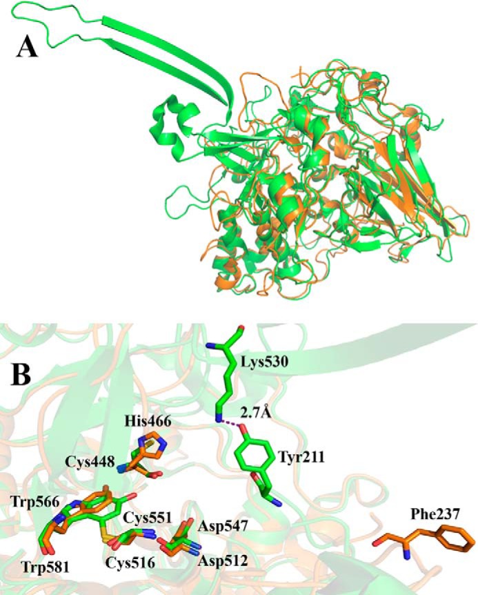
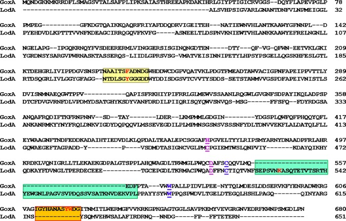
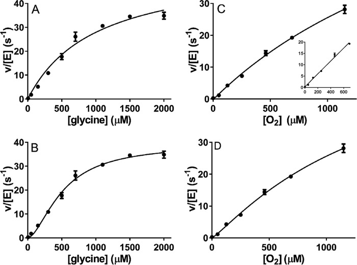
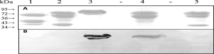
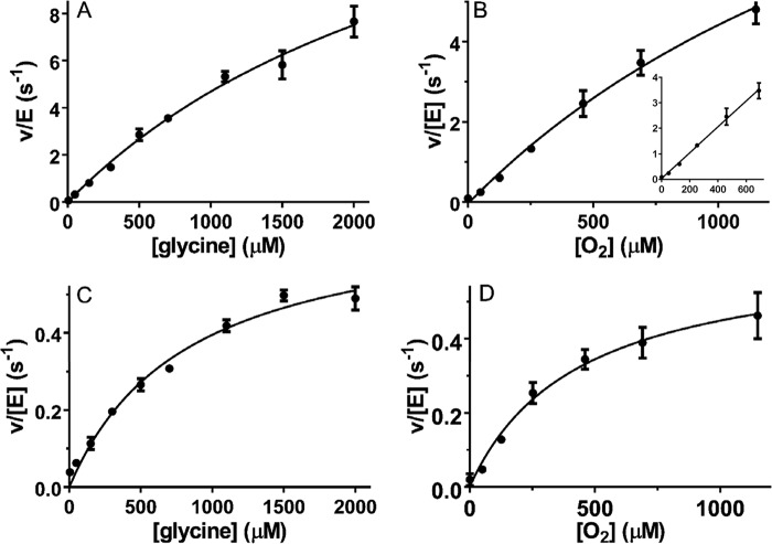
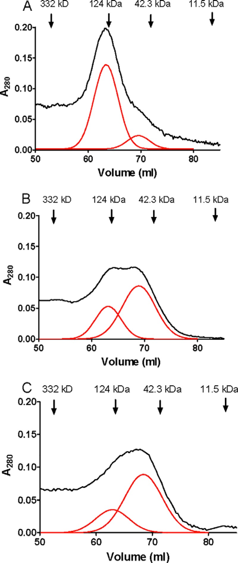
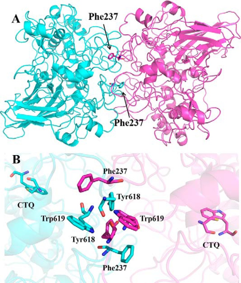
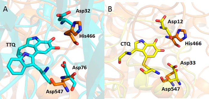
GoxA is a glycine oxidase that possesses a cysteine tryptophylquinone (CTQ) cofactor that is formed by posttranslational modifications that are catalyzed by a modifying enzyme GoxB. It is the second known tryptophylquinone enzyme to function as an oxidase, the other being the lysine ϵ-oxidase, LodA. All other enzymes containing CTQ or tryptophan tryptophylquinone (TTQ) cofactors are dehydrogenases. Kinetic analysis of GoxA revealed allosteric cooperativity for its glycine substrate, but not O2 This is the first CTQ- or TTQ-dependent enzyme to exhibit cooperativity. Here, we show that cooperativity and homodimer stabilization are strongly dependent on the presence of Phe-237. Conversion of this residue, which is a Tyr in LodA, to Tyr or Ala eliminates the cooperativity and destabilizes the dimer. These mutations also significantly affect the kcat and Km values for the substrates. On the basis of structural and modeling studies, a mechanism by which Phe-237 exerts this influence is presented. Two active site residues, Asp-547 and His-466, were also examined and shown by site-directed mutagenesis to be critical for CTQ biogenesis. This result is compared with the results of similar studies of mutagenesis of structurally conserved residues of other tryptophylquinone enzymes. These results provide insight into the roles of specific active-site residues in catalysis and CTQ biogenesis, as well as describing an interesting mechanism by which a single residue can dictate whether or not an enzyme exhibits cooperative allosteric behavior toward a substrate.

Keywords: cooperativity; enzyme kinetics; enzyme processing; oxidase; protein self-assembly; protein structure-function; quinoprotein.


Figures

FIGURE 1.
FIGURE 1.
The homology model of GoxA superimposed with the crystal structure of LodA. A, the overall structures of monomers of LodA (green) and GoxA (orange) are superimposed. B, the active sites of GoxA and LodA are superimposed. The Cys and Trp residues, which form CTQ, and selected other residues are shown as sticks. Carbons on residues of LodA and GoxA are colored green and orange, respectively. Oxygen is colored red, nitrogen is blue, and sulfur is yellow. The model was generated using the sequence of the goxA gene (Marme_1655) and the crystal structure of LodA (PDB ID 3WEU) (9).
FIGURE 2.
FIGURE 2.
Structure-based sequence alignment GoxA and LodA. The alignment of sequences is based on the homology model in Fig. 1. The Cys and Trp residues, which form CTQ, are in blue. The conserved residues, Cys-448 and Asp-512 of LodA and His-466 and Asp-547 of GoxA, are in purple. The loop that contains Phe-237 of GoxA and Tyr-211 of LodA is shaded yellow. The β-hairpin structural feature in LodA that contains Lys-530 and that is absent in GoxA is shaded green. The loop that contains Tyr-618 and Trp-619 in GoxA and that is absent in LodA is shaded orange. Key residues are in red.
FIGURE 3.
FIGURE 3.
Steady-state kinetic analysis of GoxA. A–D, assays of glycine oxidase activity were performed with varied concentrations of glycine in the presence of a fixed concentration of 1150 μm O2 (A and B) or with varied concentrations of O2 in the presence of a fixed concentration of 5 mm glycine (C and D). The lines are fits of each data set by either Equation 1 (A and C) or Equation 2 (B and D). The inset in C is a fit to a straight line of the data set with the last point omitted. Each data point was the average of a minimum of two replicates with error bars shown. In some cases, the error bars are not evident because the values were so similar that the bars are obscured by the data point.
FIGURE 4.
FIGURE 4.
SDS-PAGE and quinoprotein staining of protein fractions. A, 10% gel stained for protein. B, electroblot stained for quinoproteins. A gel identical to that in A was subjected to electrophoretic transfer of the proteins and stained for quinoproteins as described under “Experimental Procedures”. The positions of migration of molecular mass markers in the stained gel are indicated. Lane 1, D547A GoxA as isolated in complex with GoxB. Lane 2, H466A GoxA as isolated in complex with GoxB. Lane 3, WT LodA. Lane 4, active WT GoxA; Lane 5, inactive WT GoxA in complex with GoxB. The molecular masses of the proteins of interest are: LodA, 82,905 kDa; GoxA, 76,285 kDa; and GoxB, 41,856 kDa.
FIGURE 5.
FIGURE 5.
Steady-state kinetic analysis of GoxA variants. A and B, assays of the glycine oxidase activity of F237Y GoxA were performed with varied concentrations of glycine in the presence of a fixed concentration of 1150 μm O2 (A) or with varied concentrations of O2 in the presence of a fixed concentration of 5 mm glycine (B). C and D, assays of the glycine oxidase activity of F237A GoxA were performed with varied concentrations of glycine in the presence of a fixed concentration of 1150 μm O2 (C) or with varied concentrations of O2 in the presence of a fixed concentration of 5 mm glycine (D). The lines are fits to each data set by Equation 1. The inset in B is a fit to a straight line of the data set with the last point omitted. Each data point was the average of a minimum of two replicates with error bars shown. In some cases, the error bars are not evident because the values were so similar that the bars are obscured by the data point.
FIGURE 6.
FIGURE 6.
Size exclusion chromatography of WT GoxA and GoxA variants. A, WT GoxA. B, F237Y GoxA. C, F237A GoxA. The positions of the elution of the molecular mass marker proteins are indicated. The raw data are shown in black. Gaussian fits of the chromatogram that resolve overlapping peaks in the chromatogram are shown in red.
FIGURE 7.
FIGURE 7.
Docking model of the structure of the GoxA homodimer. The two monomers of GoxA are colored blue and pink. A, the overall structure of the dimer with residue Phe-237 shown as sticks. B, an expanded view of the protein-protein interface of the two monomers. The Cys and Trp residues, which form CTQ, and selected residues at the interface are shown as sticks. Carbons on residues of GoxA are colored blue on one subunit and pink on the other subunit. Oxygen is colored red, nitrogen is blue, and sulfur is yellow.
FIGURE 8.
FIGURE 8.
Residues in the active sites of MADH and QHNDH that correspond to Asp-547 and His-466 of GoxA. A, overlay of the active sites of the GoxA homology model and the MADH β-subunit (PDB ID 2BBK). The conserved active-site residues are labeled as sticks. Carbons on residues of MADH and GoxA are colored cyan and orange, respectively. B, overlay of the active sites of the GoxA homology model and the QHNDH γ-subunit (PDB ID 1JJU). Carbons on residues of QHNDH are colored yellow. Oxygen is colored red, nitrogen is blue, and sulfur is yellow.

References

    1. Campillo-Brocal J. C., Lucas-Elio P., and Sanchez-Amat A. (2013) Identification in Marinomonas mediterranea of a novel quinoprotein with glycine oxidase activity. Microbiologyopen 2, 684–694 - PMC - PubMed
    1. Davidson V. L. (2007) Protein-derived cofactors. Expanding the scope of post-translational modifications. Biochemistry 46, 5283–5292 - PubMed
    1. Davidson V. L. (2011) Generation of protein-derived redox cofactors by posttranslational modification. Mol. Biosyst. 7, 29–37 - PubMed
    1. Gómez D., Lucas-Elío P., Sanchez-Amat A., and Solano F. (2006) A novel type of lysine oxidase: l-lysine-ϵ-oxidase. Biochim. Biophys. Acta 1764, 1577–1585 - PubMed
    1. Gómez D., Lucas-Elío P., Solano F., and Sanchez-Amat A. (2010) Both genes in the Marinomonas mediterranea lodAB operon are required for the expression of the antimicrobial protein lysine oxidase. Mol. Microbiol. 75, 462–473 - PubMed

Publication types

LinkOut - more resources