Review
. 2016 Oct;39(10):656-667.
doi: 10.1016/j.tins.2016.08.004. Epub 2016 Sep 13.

Homeostatic Plasticity of Subcellular Neuronal Structures: From Inputs to Outputs

Affiliations
Review

Homeostatic Plasticity of Subcellular Neuronal Structures: From Inputs to Outputs

Winnie Wefelmeyer et al. Trends Neurosci. 2016 Oct.

Abstract

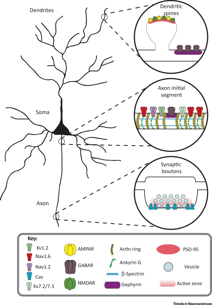
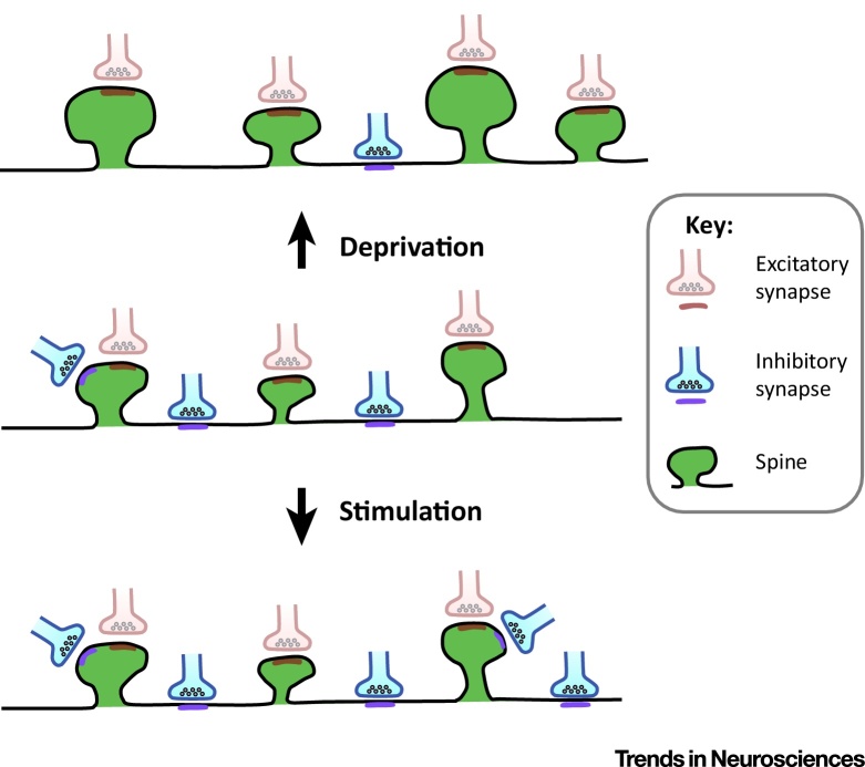
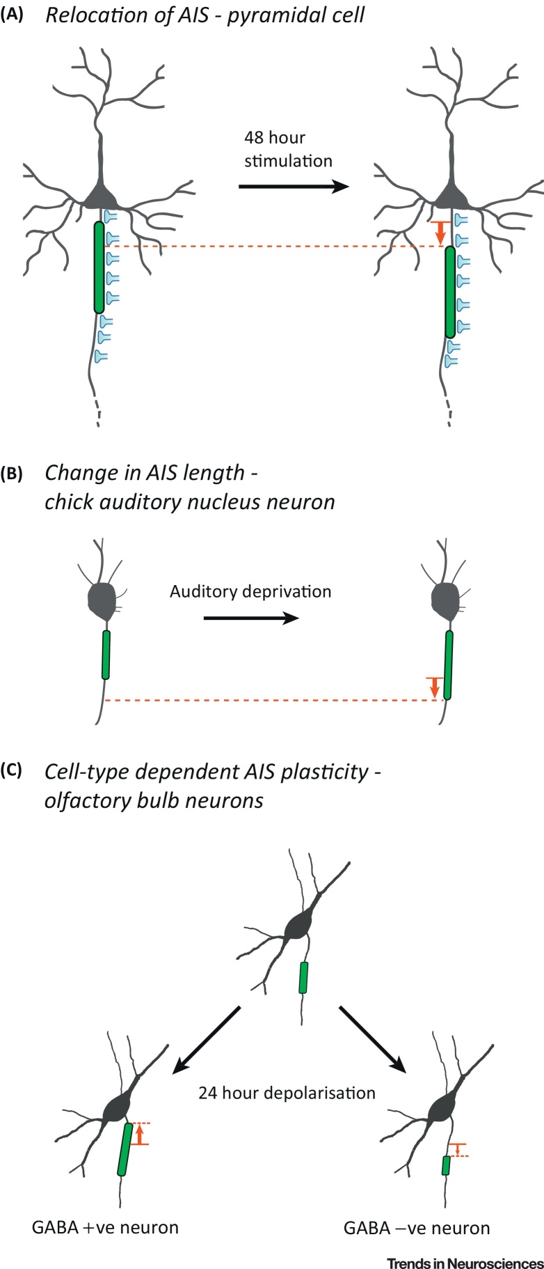
Neurons in the brain are highly plastic, allowing an organism to learn and adapt to its environment. However, this ongoing plasticity is also inherently unstable, potentially leading to aberrant levels of circuit activity. Homeostatic forms of plasticity are thought to provide a means of controlling neuronal activity by avoiding extremes and allowing network stability. Recent work has shown that many of these homeostatic modifications change the structure of subcellular neuronal compartments, ranging from changes to synaptic inputs at both excitatory and inhibitory compartments to modulation of neuronal output through changes at the axon initial segment (AIS) and presynaptic terminals. Here we review these different forms of structural plasticity in neurons and the effects they may have on network function.

Keywords: axon initial segment; dendritic spines; homeostatic plasticity; presynaptic terminals; structural plasticity.


Figures

Figure 1
Figure 1
Structural Homeostatic Plasticity Occurs at Three Main Loci in Neurons. Inputs arrive primarily at the dendrites of neurons (top right), where synapses are formed on dendritic spines and the dendritic shaft. AMPA and NMDA receptors are located mainly in spines while inhibitory GABAA receptors are found mostly on the dendritic shaft. At the axon initial segment (AIS) (middle right), ankyrin G tethers a large complement of voltage-gated channels, including NaV1.2, NaV1.6, Kv7.2, and Kv7.3, to the membrane. Inhibitory synapses are found localised to gephyrin in gaps between ankyrin G that also contain KV1.2. These components at the AIS initiate and shape neuronal output, which is transmitted along the axon. At presynaptic boutons (bottom right), activation of voltage-gated calcium channels by the action potential leads to exocytosis of neurotransmitter-filled vesicles at the active zone and thus transmission of the neuron's output signal.
Figure 2
Figure 2
Inhibitory Synapses Formed on Pyramidal Cell Dendrites Show Homeostatic Plasticity in Response to Modulation of Activity Levels. At rest (middle), inhibitory boutons synapse mainly onto the dendritic shaft, although some innervate dendritic spines . After sensory deprivation (top), inhibitory synapses are removed from both the dendritic shaft and the spines , , . At the same time, spine size and density increase , . Conversely, after long-term sensory stimulation, the density of inhibitory innervation on both shafts and spines increases (bottom).
Figure 3
Figure 3
Cell Type-Specific Structural Plasticity of the Axon Initial Segment (AIS). (A) Pyramidal cells move their AIS away from the soma after long-term stimulation , , , . Importantly, GABAergic axoaxonic inputs to the AIS do not change, creating a partial mismatch between the two compartments and an overall decrease in excitability . (B) Auditory deprivation increases the length of the AIS in chick auditory nucleus neurons resulting in an increase in excitability . (C) Following 24 h of depolarisation, GABAergic olfactory bulb interneurons show proximal lengthening of their AIS, which brings it closer to the soma. By contrast, neighbouring non-GABAergic interneurons respond by shortening their AIS instead, resulting in a more-distal start position .

References

    1. Cajal S.R.Y., Swanson N. 1st edn. Oxford University Press; 1995. Histology of the Nervous System of Man and Vertebrates.
    1. Scott E.K., Luo L. How do dendrites take their shape? Nat. Neurosci. 2001;4:359–365. - PubMed
    1. Debanne D. Information processing in the axon. Nat. Rev. Neurosci. 2004;5:304–316. - PubMed
    1. Chklovskii D.B. Synaptic connectivity and neuronal morphology: two sides of the same coin. Neuron. 2004;43:609–617. - PubMed
    1. Holtmaat A., Svoboda K. Experience-dependent structural synaptic plasticity in the mammalian brain. Nat. Rev. Neurosci. 2009;10:647–658. - PubMed

LinkOut - more resources