. 2016 Sep 15;9(2):340-352.
doi: 10.18632/aging.101035.

Protective effects of alpha phenyl-tert-butyl nitrone and ascorbic acid in human adipose derived mesenchymal stem cells from differently aged donors

Affiliations

Protective effects of alpha phenyl-tert-butyl nitrone and ascorbic acid in human adipose derived mesenchymal stem cells from differently aged donors

Adiv A Johnson et al. Aging (Albany NY). .

Abstract

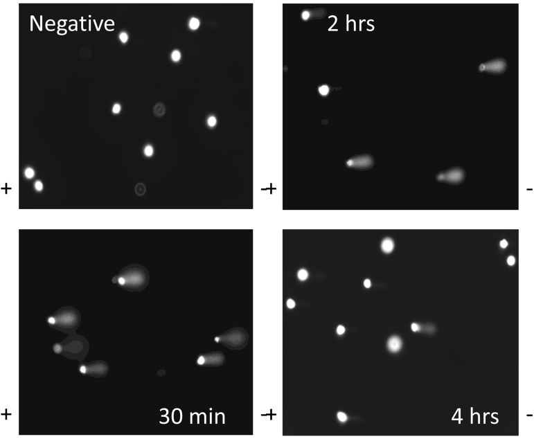
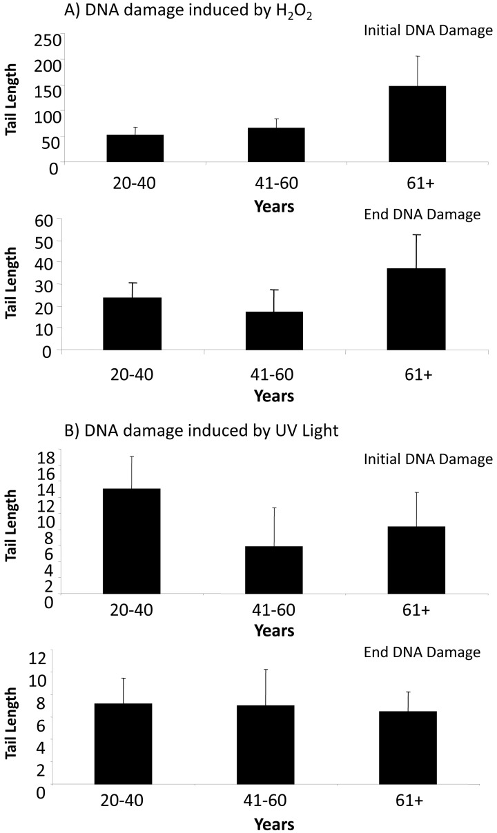
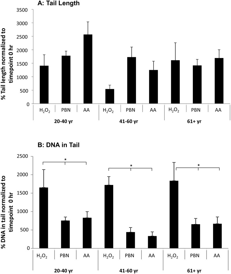
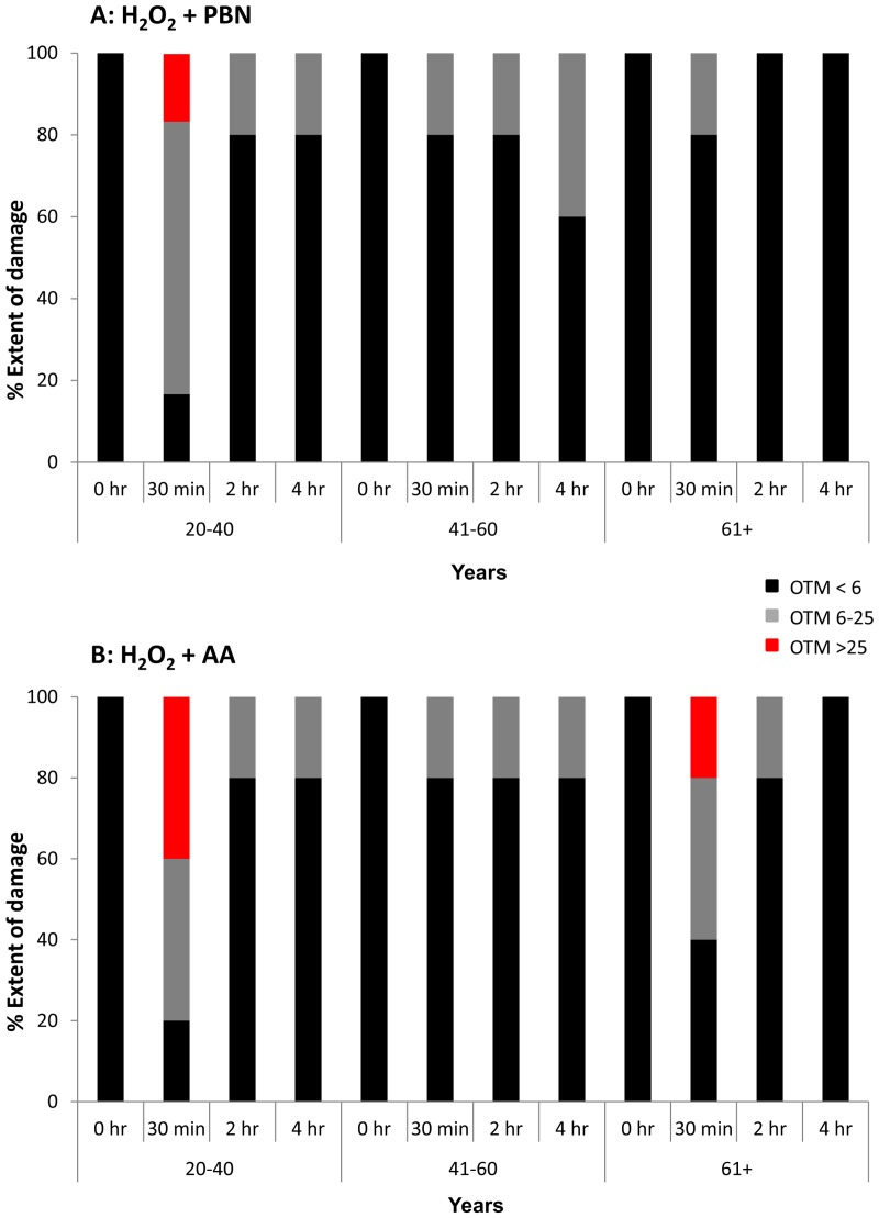
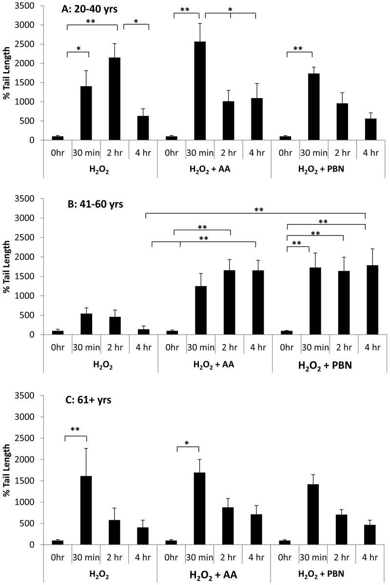
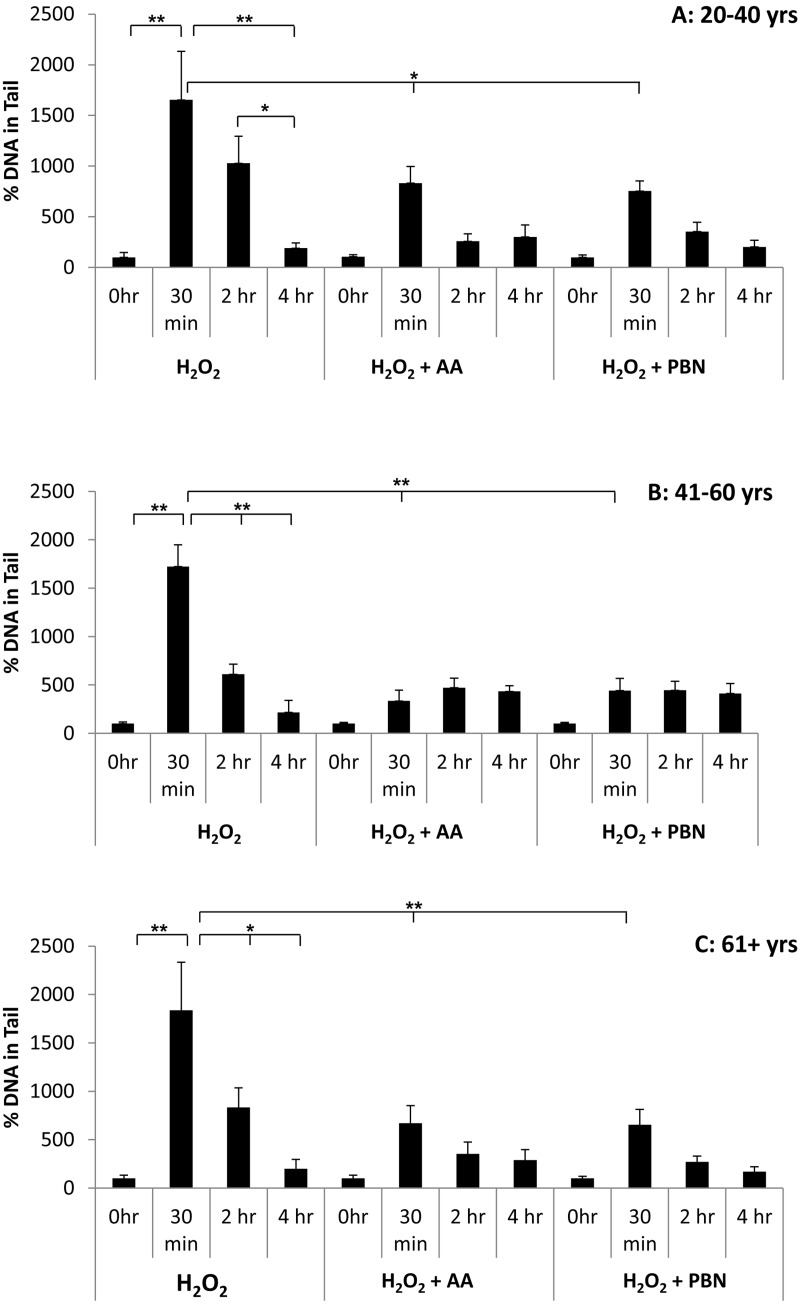
Adipose-derived mesenchymal stem cells (ADSCs) are multipotent stem cells that promote therapeutic effects and are frequently used in autologous applications. Little is known about how ADSCs respond to genotoxic stress and whether or not donor age affects DNA damage and repair. In this study, we used the comet assay to assess DNA damage and repair in human ADSCs derived from young (20-40 years), middle-aged (41-60 years), and older (61+ years) donors following treatment with H2O2 or UV light. Tail lengths in H2O2-treated ADSCs were substantially higher than the tail lengths in UV-treated ADSCs. After 30 minutes of treatment with H2O2, ADSCs preconditioned with alpha phenyl-tert-butyl nitrone (PBN) or ascorbic acid (AA) showed a significant reduction in % tail DNA. The majority of ADSCs treated with PBN or AA displayed low olive tail movements at various timepoints. In general and indicative of DNA repair, % tail length and % tail DNA peaked at 30 minutes and then decreased to near-control levels at the 2 hour and 4 hour timepoints. Differently aged ADSCs displayed comparable levels of DNA damage in the majority of these experiments, suggesting that the age of the donor does not affect the DNA damage response in cultured ADSCs.

Keywords: ADSCs; DNA damage; DNA repair; aging; antioxidants; comet assay.


Conflict of interest statement

CONFLICTS OF INTEREST

The authors declare that they have no conflicts of interest.

Figures

Figure 1
Figure 1. DNA comets after treatment of ADSCs with H2O2
Representative slides of DNA comets from human ADSCs treated with H2O2 for 30 minutes are shown. Slides are shown for ADSCs untreated with H2O2 (negative control) or H2O2-treated ADSCs at the 30 minute, 2 hour, and 4 hour timepoints. DNA comet tails were notably pronounced at the 30 minute and 2 hour timepoints. The comet tails decreased in length at the 4 hour timepoint.
Figure 2
Figure 2. DNA damage in ADSCs treated with H2O2 or UV light
(A) Tail length was quantified in ADSCs derived from young (20-40 years), middle-aged (41-60 years), and older (61+ years) after treatment with H2O2 for 30 minutes. Tail length was measured at the 30 minute (initial DNA damage) and 4 hour (end DNA damage) timepoints. Although tail length was longer in the ADSCs derived from 61+ year old donors, the tail length in differently aged ADSCs was statistically comparable. (B) Tail length was measured in ADSCs from differently aged sources following treatment with UV light for 30 minutes. Tail length was statistically comparable in ADSCs derived from young, middle-aged, and older donors. In comparison to ADSCs treated with H2O2, tail length was drastically shorter in ADSCs treated with UV light.
Figure 3
Figure 3. PBN and AA reduce DNA damage in H2O2-treated ADSCs
ADSCs from young, middle-aged, and older sources were preincubated for 24 hours with either PBN or AA and then treated with H2O2 for 30 minutes. (A) ADSCs preincubated with either PBN or AA displayed comparable % tail lengths to the controls, which were treated with H2O2 alone and not preincubated with PBN or AA. (B) ADSCs preincubated with either PBN or AA showed a significant reduction in % tail DNA compared to the controls. This was true for ADSCs derived from each age group. * indicates p < 0.05.
Figure 4
Figure 4. OTM was low in the majority of H2O2-treated ADSCs preincubated with PBN or AA
Olive tail movement (OTM) at the 0 hour, 30 minute, 2 hour, and 4 hour timepoints was measured in H2O2-treated ADSCs additionally treated with PBN or AA. The % of the ADSC population showing an OTM < 6 (black), an OTM between 6 and 25 (grey), or an OTM > 25 (red) is shown at each timepoint. (A) In general, the OTM was quite low (< 6) in ADSCs treated with PBN. Higher OTMs were found at the 30 minute timepoint in ADSCs derived from 20-40 year old donors. (B) Similarly, the majority of ADSCs treated with AA showed an OTM < 6. Higher OTMs were observed at the 30 minute timepoint in ADSCs derived from 20-40 year old and 61+ year old donors.
Figure 5
Figure 5. Changes in % tail length over time in ADSCs treated with H2O2
% tail length was measured in ADSCs derived from 20-40 year old donors (A), 41-60 year old donors (B), or 61+ year old donors (C) at the 0 hour, 30 minute, 2 hour, and 4 hour timepoints after treatment with H2O2 for 30 minutes. % tail length was measured in ADCSs treated with H2O2 alone or in ADSCs additionally treated with AA or PBN. * indicates p < 0.05 and ** indicates p < 0.005.
Figure 6
Figure 6. Changes in % tail DNA over time in ADSCs treated with H2O2
% tail DNA was measured in ADSCs derived from 20-40 year old donors (A), 41-60 year old donors (B), or 61+ year old donors (C) at the 0 hour, 30 minute, 2 hour, and 4 hour timepoints after treatment with H2O2 for 30 minutes. % tail DNA was measured in ADCSs treated with H2O2 alone or in ADSCs additionally treated with AA or PBN. * indicates p < 0.05 and ** indicates p < 0.005.

References

    1. Minteer D, Marra KG, Rubin JP. Adipose-derived mesenchymal stem cells: biology and potential applications. Adv Biochem Eng Biotechnol. 2013;129:59–71. doi: 10.1007/10_2012_146. - DOI - PubMed
    1. Navarro-Betancourt JR, Hernández S. On the existence of cardiomesenchymal stem cells. Med Hypotheses. 2015;84:511–15. doi: 10.1016/j.mehy.2015.02.011. - DOI - PubMed
    1. Alonso-Alonso ML, Srivastava GK. Current focus of stem cell application in retinal repair. World J Stem Cells. 2015;7:641–48. doi: 10.4252/wjsc.v7.i3.641. - DOI - PMC - PubMed
    1. Ra JC, Kang SK, Shin IS, Park HG, Joo SA, Kim JG, Kang BC, Lee YS, Nakama K, Piao M, Sohl B, Kurtz A. Stem cell treatment for patients with autoimmune disease by systemic infusion of culture-expanded autologous adipose tissue derived mesenchymal stem cells. J Transl Med. 2011;9:181. - PMC - PubMed
    1. Sowa Y, Imura T, Numajiri T, Nishino K, Fushiki S. Adipose-derived stem cells produce factors enhancing peripheral nerve regeneration: influence of age and anatomic site of origin. Stem Cells Dev. 2012;21:1852–62. doi: 10.1089/scd.2011.0403. - DOI - PubMed