Review
. 2016 Aug;231(1):73-84.
doi: 10.1086/689592.

Imaging Adenosine Triphosphate (ATP)

Affiliations
Review

Imaging Adenosine Triphosphate (ATP)

Megha Rajendran et al. Biol Bull. 2016 Aug.

Abstract

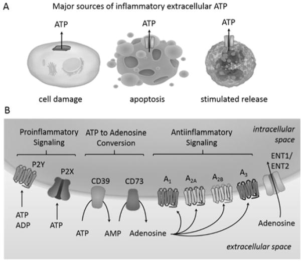
Adenosine triphosphate (ATP) is a universal mediator of metabolism and signaling across unicellular and multicellular species. There is a fundamental interdependence between the dynamics of ATP and the physiology that occurs inside and outside the cell. Characterizing and understanding ATP dynamics provide valuable mechanistic insight into processes that range from neurotransmission to the chemotaxis of immune cells. Therefore, we require the methodology to interrogate both temporal and spatial components of ATP dynamics from the subcellular to the organismal levels in live specimens. Over the last several decades, a number of molecular probes that are specific to ATP have been developed. These probes have been combined with imaging approaches, particularly optical microscopy, to enable qualitative and quantitative detection of this critical molecule. In this review, we survey current examples of technologies available for visualizing ATP in living cells, and identify areas where new tools and approaches are needed to expand our capabilities.


Figures

Figure 1
Figure 1
Extracellular ATP. (A) Mechanical damage of the cell membrane can cause uncontrolled ATP release whereas ATP release from apoptotic or inflammatory cells is mediated by transmembrane channels. (B) Extracellular ATP and ADP act as signaling molecules by binding P2 receptors, which are classified into 2 families based on their structure. P2Y receptors are GPCRs whereas P2X receptors are nucleotide-gated ion channels. Activation of P2Rs is generally proinflammatory. Extracellular ATP and ADP are converted to adenosine by the action of the ectonucleotidases CD39 and CD73. Adenosine signaling can occur through four structurally related GPCRs and generally leads to decreased inflammation. Adenosine is cleared from the extracellular space by the equilibrative nucleoside transporters 1 and 2 (ENT1, ENT2).
Figure 2
Figure 2
Tools for ATP Visualization. (A) Quinacrine is a fluorescent dye that can stain vesicular ATP. (B) Mant-ATP is a fluorescent analogue. (C) Nanoflare using an ATP aptamer attached to a gold nanoparticle. In the absence of ATP, a competitor strand that is conjugated to a fluorescent dye is bound to the aptamer, but fluorescence is quenched by proximity to the gold. When ATP binds, the competitor strand is released and fluorescence is de-quenched. (D) Luciferase chemiluminescence requires a luciferin substrate and ATP. (E) The ATeam sensors use the F1F0 ATP synthase ε subunit as an ATP binding domain attached to a FRET pair of fluorescent proteins. (F) The Perceval sensors use the PII family protein GlnK as an ATP binding domain attached to a circularly permuted fluorescent protein. For (A) – (D) the sensor or a substrate must be exogenously supplied and loaded into cells or tissue. For (E) – (F) the sensors are completely genetically-encoded.

References

    1. Ainscow EK, Mirshamsi S, Tang T, Ashford MLJ, Rutter GA. Dynamic imaging of free cytosolic ATP concentration during fuela sensing by rat hypothalamic neurones: evidence for ATP-independent control of ATP-sensitive K+ channels. J Physiol. 2002;544:429–445. - PMC - PubMed
    1. Akopova I, Tatur S, Grygorczyk M, Luchowski R, Gryczynski I, Gryczynski Z, Borejdo J, Grygorczyk R. Imaging exocytosis of ATP-containing vesicles with TIRF microscopy in lung epithelial A549 cells. Purinergic Signal. 2012;8:59–70. - PMC - PubMed
    1. Befroy DE, Rothman DL, Petersen KF, Shulman GI. 31P-magnetization transfer magnetic resonance spectroscopy measurements of in vivo metabolism. Diabetes. 2012;61:2669–2678. - PMC - PubMed
    1. Bélanger M, Allaman I, Magistretti PJ. Brain energy metabolism: focus on astrocyte-neuron metabolic cooperation. Cell Metab. 2011;14:724–738. - PubMed
    1. Bell CJ, Manfredi G, Griffiths EJ, Rutter GA. Luciferase expression for ATP imaging: application to cardiac myocytes. Methods Cell Biol. 2007;80:341–352. - PubMed

Publication types

MeSH terms