

Functional segregation and integration within fronto-parietal networks
- PMID: 27639357
- PMCID: PMC5312783
- DOI: 10.1016/j.neuroimage.2016.08.031
Functional segregation and integration within fronto-parietal networks
Abstract
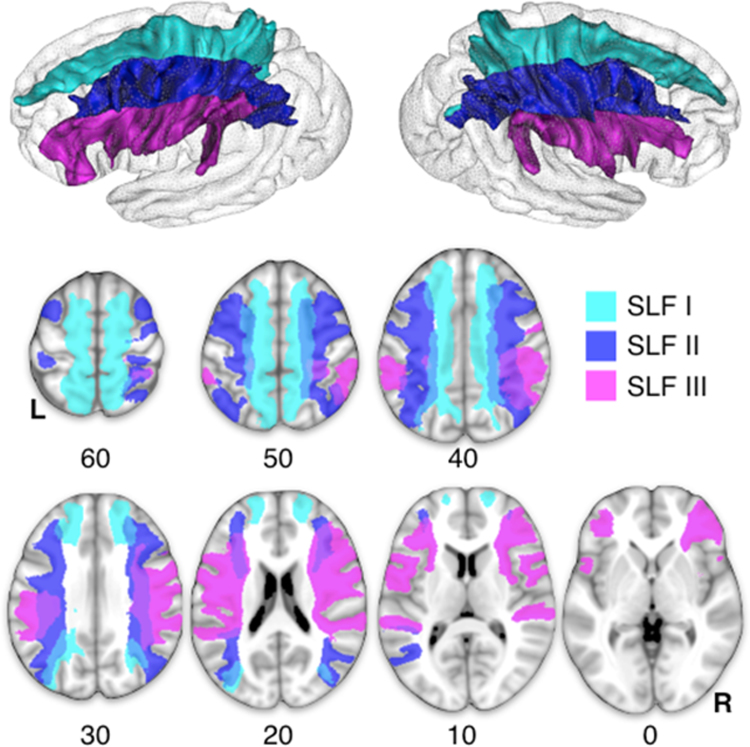
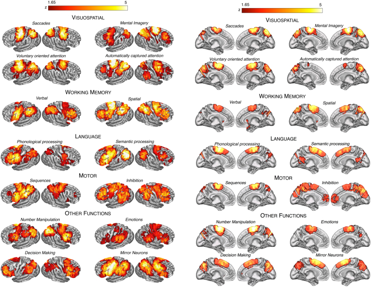
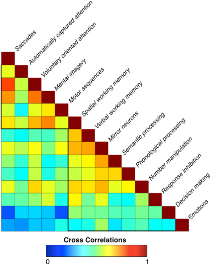
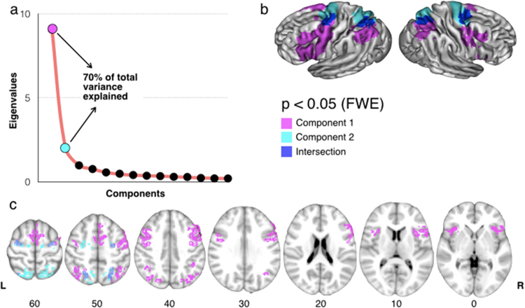
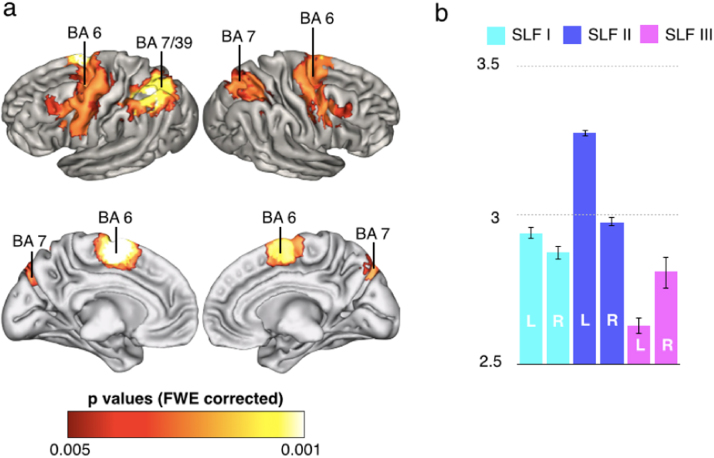
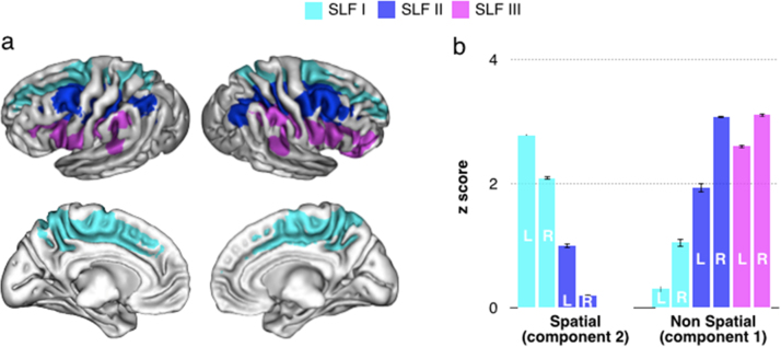
Experimental data on monkeys and functional studies in humans support the existence of a complex fronto-parietal system activating for cognitive and motor tasks, which may be anatomically supported by the superior longitudinal fasciculus (SLF). Advanced tractography methods have recently allowed the separation of the three branches of the SLF but are not suitable for their functional investigation. In order to gather comprehensive information about the functional organisation of these fronto-parietal connections, we used an innovative method, which combined tractography of the SLF in the largest dataset so far (129 participants) with 14 meta-analyses of functional magnetic resonance imaging (fMRI) studies. We found that frontal and parietal functions can be clustered into a dorsal spatial/motor network associated with the SLF I, and a ventral non-spatial/motor network associated with the SLF III. Further, all the investigated functions activated a middle network mostly associated with the SLF II. Our findings suggest that dorsal and ventral fronto-parietal networks are segregated but also share regions of activation, which may support flexible response properties or conscious processing. In sum, our novel combined approach provided novel findings on the functional organisation of fronto-parietal networks, and may be successfully applied to other brain connections.
Keywords: Diffusion tractography; Frontal parietal; Functional magnetic resonance imaging (fMRI); Meta-analysis; Superior longitudinal fasciculus.
Copyright © 2016 The Authors. Published by Elsevier Inc. All rights reserved.
Figures
References
-
- Abhinav K., Pathak S., Richardson R.M., Engh J., Gardner P., Yeh F.C., Friedlander R.M., Fernandez-Miranda J.C. Application of high-definition fiber tractography in the management of supratentorial cavernous malformations: a combined qualitative and quantitative approach. Neurosurgery. 2014;74:668–680. Discussion 680-661. - PubMed
-
- Agosta F., Galantucci S., Canu E., Cappa S.F., Magnani G., Franceschi M., Falini A., Comi G., Filippi M. Disruption of structural connectivity along the dorsal and ventral language pathways in patients with nonfluent and semantic variant primary progressive aphasia: a DT MRI study and a literature review. Brain Lang. 2013;127:157–166. - PubMed
-
- Avants, B.B., Duda, J.T., Zhang, H., Gee, J.C., 2007. Multivariate normalization with symmetric diffeomorphisms for multivariate studies. Medical image computing and computer-assisted intervention. In: Proceedings of the MICCAI International Conference on Medical Image Computing and Computer-Assisted Intervention 10. pp. 359–366. - PubMed
-
- Badre D., D’Esposito M. Functional magnetic resonance imaging evidence for a hierarchical organization of the prefrontal cortex. J. Cogn. Neurosci. 2007;19:2082–2099. - PubMed
Publication types
MeSH terms
Grants and funding
LinkOut - more resources
Full Text Sources
Other Literature Sources
Molecular Biology Databases
