

Total average diastolic longitudinal displacement by colour tissue doppler imaging as an assessment of diastolic function
- PMID: 27639377
- PMCID: PMC5027100
- DOI: 10.1186/s12947-016-0083-2
Total average diastolic longitudinal displacement by colour tissue doppler imaging as an assessment of diastolic function
Abstract
Background: The current method for a non-invasive assessment of diastolic dysfunction is complex with the use of algorithms of many different echocardiographic parameters. Total average diastolic longitudinal displacement (LD), determined by colour tissue Doppler imaging (TDI) via the measurement of LD during early diastole and atrial contraction, can potentially be used as a simple and reliable alternative.
Methods: In 206 patients, using GE Healthcare Vivid E7 and 9 and Echopac BT11 software, we determined both diastolic LD, measured in the septal and lateral walls in the apical 4-chamber view by TDI, and the degree of diastolic dysfunction, based on current guidelines. Of these 206 patients, 157 had cardiac anomalies that could potentially affect diastolic LD such as severe systolic heart failure (n = 45), LV hypertrophy (n = 49), left ventricular (LV) dilation (n = 30), and mitral regurgitation (n = 33). Intra and interobserver variability of diastolic LD measures was tested in 125 patients.
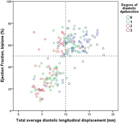
Results: A linear relationship between total average diastolic LD and the degree of diastolic dysfunction was found. A total average diastolic LD of 10 mm was found to be a consistent threshold for the general discrimination of patients with or without diastolic dysfunction. Using linear regression, total average diastolic LD was estimated to fall by 2.4 mm for every increase in graded severity of diastolic dysfunction (β = -0.61, p-value <0.001). Patients with LV hypertrophy had preserved total average diastolic LD despite being classified as having diastolic dysfunction. Reproducibility of LD measures was acceptable.
Conclusions: There is strong evidence suggesting that patients with a total average diastolic LD under 10 mm have diastolic dysfunction.
Keywords: Colour tissue Doppler imaging; Diastolic function; Diastolic longitudinal displacement; Echocardiography.
Figures


References
-
- Nagueh SF, Smiseth OA, Appleton CP, Byrd BF, Dokainish H, Edvardsen T, et al. Recommendations for the evaluation of left ventricular diastolic function by echocardiography: an update from the American society of echocardiography and the European association of cardiovascular imaging. J Am Soc Echocardiogr. 2016;29(4):277–314. doi: 10.1016/j.echo.2016.01.011. - DOI - PubMed
-
- Petrie MC, Hogg K, Caruana L, McMurray JJV. Poor concordance of commonly used echocardiographic measures of left ventricular diastolic function in patients with suspected heart failure but preserved systolic function: is there a reliable echocardiographic measure of diastolic dysfunction? Heart. 2004;90(5):511–7. doi: 10.1136/hrt.2003.011403. - DOI - PMC - PubMed
-
- Paulus WJ, Tschöpe C, Sanderson JE, Rusconi C, Flachskampf FA, Rademakers FE, et al. How to diagnose diastolic heart failure: a consensus statement on the diagnosis of heart failure with normal left ventricular ejection fraction by the Heart Failure and Echocardiography Associations of the European Society of Cardiology. Eur Heart J. 2007;28(20):2539–50. doi: 10.1093/eurheartj/ehm037. - DOI - PubMed
MeSH terms
LinkOut - more resources
Full Text Sources
Other Literature Sources
Research Materials
