

Linked color imaging application for improving the endoscopic diagnosis accuracy: a pilot study
- PMID: 27641243
- PMCID: PMC5027569
- DOI: 10.1038/srep33473
Linked color imaging application for improving the endoscopic diagnosis accuracy: a pilot study
Abstract
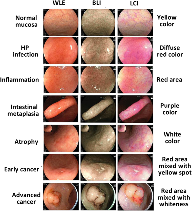
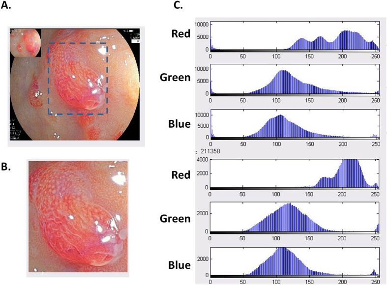
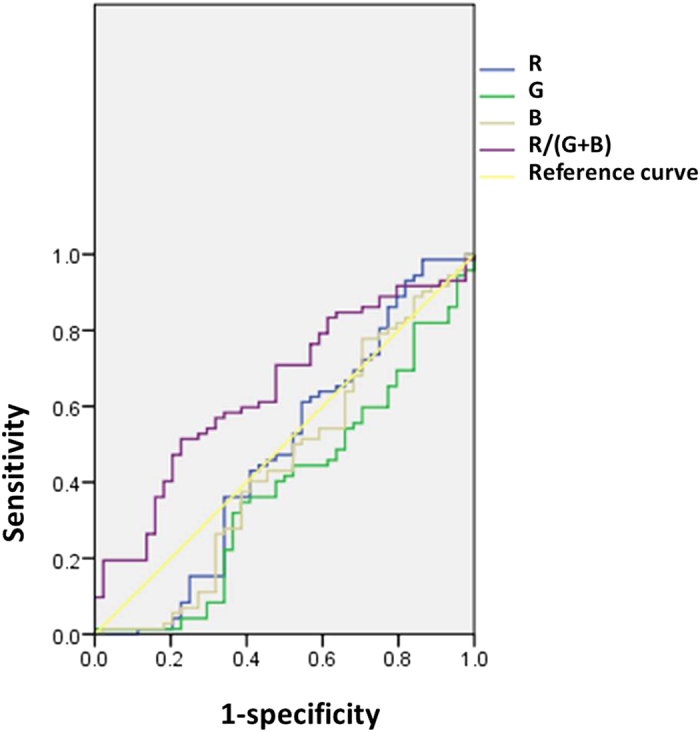
Endoscopy has been widely used in diagnosing gastrointestinal mucosal lesions. However, there are still lack of objective endoscopic criteria. Linked color imaging (LCI) is newly developed endoscopic technique which enhances color contrast. Thus, we investigated the clinical application of LCI and further analyzed pixel brightness for RGB color model. All the lesions were observed by white light endoscopy (WLE), LCI and blue laser imaging (BLI). Matlab software was used to calculate pixel brightness for red (R), green (G) and blue color (B). Of the endoscopic images for lesions, LCI had significantly higher R compared with BLI but higher G compared with WLE (all P < 0.05). R/(G + B) was significantly different among 3 techniques and qualified as a composite LCI marker. Our correlation analysis of endoscopic diagnosis with pathology revealed that LCI was quite consistent with pathological diagnosis (P = 0.000) and the color could predict certain kinds of lesions. ROC curve demonstrated at the cutoff of R/(G+B) = 0.646, the area under curve was 0.646, and the sensitivity and specificity was 0.514 and 0.773. Taken together, LCI could improve efficiency and accuracy of diagnosing gastrointestinal mucosal lesions and benefit target biopsy. R/(G + B) based on pixel brightness may be introduced as a objective criterion for evaluating endoscopic images.
Figures
References
-
- Dohi O. et al.. Diagnostic ability of magnifying endoscopy with blue laser imaging for early gastric cancer: a prospective study. Gastric cancer: official journal of the International Gastric Cancer Association and the Japanese Gastric Cancer Association, doi: 10.1007/s10120-016-0620-6 (2016). - DOI - PubMed
MeSH terms
LinkOut - more resources
Full Text Sources
Other Literature Sources
Medical
