

doi: 10.1186/s13567-016-0375-4.
First case of chronic wasting disease in Europe in a Norwegian free-ranging reindeer
Affiliations
- PMID: 27641251
- PMCID: PMC5024462
- DOI: 10.1186/s13567-016-0375-4
Item in Clipboard
First case of chronic wasting disease in Europe in a Norwegian free-ranging reindeer
Vet Res.
.
Abstract
Chronic wasting disease (CWD) is a fatal contagious prion disease in cervids that is enzootic in some areas in North America. The disease has been found in deer, elk and moose in the USA and Canada, and in South Korea following the importation of infected animals. Here we report the first case of CWD in Europe, in a Norwegian free-ranging reindeer in Southern Norway. The origin of the disease is unknown. Until now a low number of cervids, and among them a few reindeer, have been tested for CWD in Norway. Therefore the prevalence of CWD is unknown.
Figures
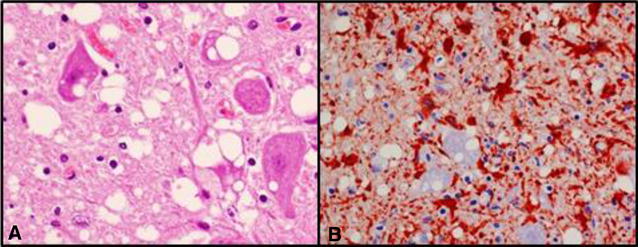
Histology of the obex. A HE staining showing vacuoles in the neurons and neuropil ×600. B Glial fibrillary acidic protein (GFAP) immunolabelling showing strong proliferation of reactive astrocytes (gliosis) in the obex area ×400.
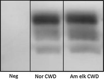
Western blot detection of PrP
res
with TeSeE Western blot kit (Bio-Rad), using SHa31 and P4 mAb. The Norwegian reindeer sample (Nor CWD) showed a typical 3-band pattern similar to the American elk CWD sample (Am elk CWD). No signal was seen in the negative sheep brain sample (Neg).
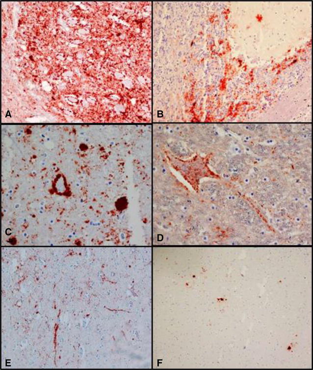
IHC labelling of PrP
CWD
using F89/160.1.5 and 2G11 mAb. A Obex, intense coarse particulate labelling in the DMNV ×200. B Cerebellum, patchy labelling in the granular layer and stellate in the molecular layer ×200. C Ventral midbrain, scattered granules or accumulations of PrPCWD that appear as plaques ×400. D Ventral midbrain, neuronal and axonal labelling ×400. E Ventral midbrain, linear type labelling ×100. F Dorsal midbrain, sparse immunolabelling, plaque-like accumulation of PrPCWD ×100.
References
-
- USGS National Wildlife Health Center. Map of chronic wasting disease in North America. http://www.nwhc.usgs.gov/disease_information/chronic_wasting_disease/. Accessed 11 Aug 2016
-
- Spraker TR, Miller MW, Williams ES, Getzy DM, Adrian WJ, Schoonveld GG, Spowart RA, O’Rourke KI, Miller JM, Merz PA. Spongiform encephalopathy in free-ranging mule deer (Odocoileus hemionus), white-tailed deer (Odocoileus virginianus) and rocky mountain elk (Cervus elaphus nelsoni) in northcentral Colorado. J Wildl Dis. 1997;33:1–6. doi: 10.7589/0090-3558-33.1.1. - DOI - PubMed
MeSH terms
LinkOut - more resources
Full Text Sources
Other Literature Sources
