. 2016 Dec 1;19(6):800-807.
doi: 10.1016/j.stem.2016.08.006. Epub 2016 Sep 15.

CD82 Is a Marker for Prospective Isolation of Human Muscle Satellite Cells and Is Linked to Muscular Dystrophies

Affiliations

CD82 Is a Marker for Prospective Isolation of Human Muscle Satellite Cells and Is Linked to Muscular Dystrophies

Matthew S Alexander et al. Cell Stem Cell. .

Abstract

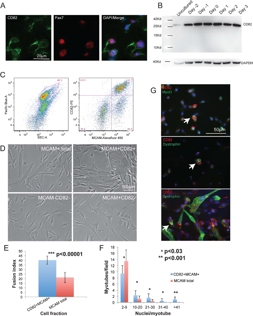
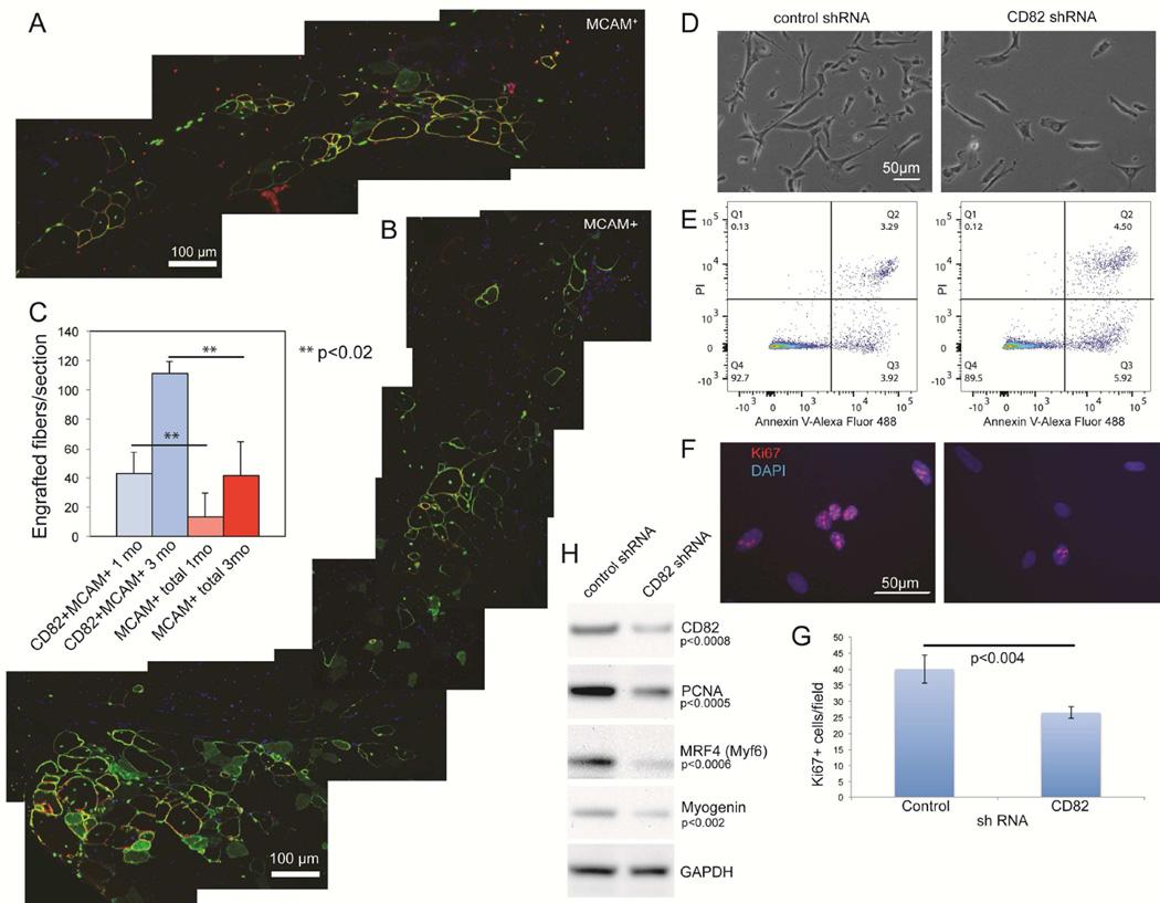
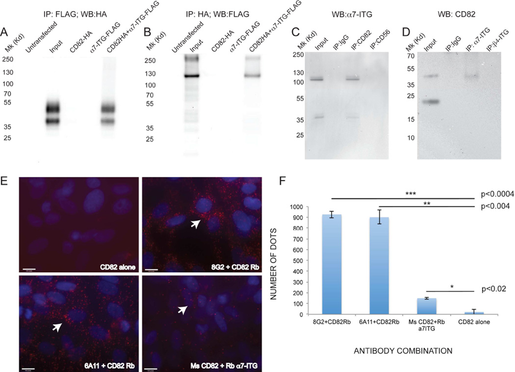
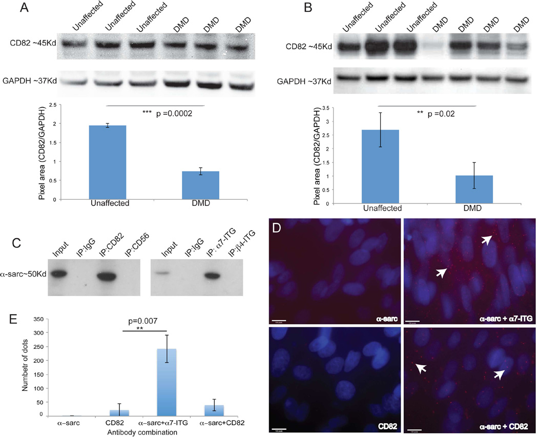
Cell-surface markers for prospective isolation of stem cells from human skeletal muscle have been difficult to identify. Such markers would be powerful tools for studying satellite cell function during homeostasis and in pathogenesis of diseases such as muscular dystrophies. In this study, we show that the tetraspanin KAI/CD82 is an excellent marker for prospectively isolating stem cells from human fetal and adult skeletal muscle. Human CD82+ muscle cells robustly engraft into a mouse model of muscular dystrophy. shRNA knockdown of CD82 in myogenic cells reduces myoblast proliferation, suggesting it is functionally involved in muscle homeostasis. CD82 physically interacts with alpha7beta1 integrin (α7β1-ITG) and with α-sarcoglycan, a member of the Dystrophin-Associated Glycoprotein Complex (DAPC), both of which have been linked to muscular dystrophies. Consistently, CD82 expression is decreased in Duchenne muscular dystrophy patients. Together, these findings suggest that CD82 function may be important for muscle stem cell function in muscular disorders.


Figures

Figure 1
Figure 1. CD82 is a prospective marker for muscle satellite cells
A) Cross sections of human fetal skeletal muscle immunostained for CD82 (green) and Pax7 (red). Co-staining confirmed that satellite cells express CD82. Nuclei are stained in blue with DAPI. Scale bars=50µm. B) Western blot of differentiating human fetal cells showing expression of CD82 (~30Kd) at all timepoints analyzed. C) FACS purification of myogenic cells from human fetal tissue using CD82 and MCAM. Live cells were gated based on Calcein Blue signal (left) and 4 cell fractions were isolated: MCAM+CD82+, MCAM+CD82, MCAMCD82 and MCAM+ total (MCAM+CD82+ and MCAM+CD82 combined). D) Cells fractions were plated at the same density and induced to differentiate for 48 hours (scale bar: 100µm). MCAMCD82 and MCAM+CD82 never fused, suggesting that selection for both MCAM and CD82 enriches for myogenic activity. E) The fusion index of MCAM+CD82+ at 48hrs following differentiation was significantly higher than in MCAM total (p<0.00001, n=5 samples). F) Myotube size is significantly increased in MCAM+CD82+ cells compared to MCAM total. For each sample, 10 independent microscopic images with 250–540 nuclei/image were analyzed. Small myotubes (2–9 nuclei in size) are significantly increased in MCAM total cells, while large myotubes containing >41 nuclei were only seen in the MCAM+CD82+ cell fraction. Data are represented as mean ± SEM and p values were calculated via t-test. G) Co-expression of CD82, MyoD and dystrophin in cultured primary human fetal muscle cells (P1). CD82 localizes to the cell membrane of mononuclear myogenic cells expressing MyoD or dystrophin (arrows) (Dumont et al., 2015) and it is maintained in differentiating cultures. Scale bar: 50 microns.
Figure 2
Figure 2. CD82+MCAM+ human muscle cells engraft in immune-deficient mice with muscular dystrophy
IM injections were performed in the tibialis anterior (TA) muscle of recipient NODRag1nullmdx5cv mice. 100,000 cells positive for MCAM alone (MCAM+) were injected in one TA, while the contralateral TA received 100,000 double-positive cells (MCAM+CD82+). A) Merged images showing the entire injection site 3 months after transplantation of human fetal MCAM+ cells. B) Merged images showing the entire injection site using human fetal MCAM+CD82+ cells 3 months following transplantation. Red: human spectrin; green: human lamin A/C (nuclei contour) and dystrophin (myofiber contour); blue: nuclei (DAPI) C) Engraftment quantification was performed based on expression of both human specific spectrin and dystrophin (Rozkalne et al., 2014). At 4 weeks after transplantation we observed significantly more myofibers of human origin in muscles injected with MCAM+CD82+cells compared to MCAM+ cells (**p<0.02). The same was true for muscles harvested 3 months after transplantation (n=7 per each group, **p<0.02). D) CD82-shRNA and control shRNA on primary human muscle cells using commercial lentiviruses (Santa Cruz Biotechnology). The total number of cells was significantly decreased in CD82-sh compared to control-sh cultures (t-test p<0.03). Apoptosis assays utilized Annexin V detection (D), since caspase 3 activity can regulate other functions, including self-renewal of satellite cells (Dick et al., 2015). No significant differences in apoptosis were seen between control- and CD82-sh cultures. Ki67 immunostaining (F) was used to determine the number of proliferating cells in the cultures, which was significantly decreased in CD82shRNA cultures via t-test (G). H) Western blot analyses in control- and CD82-shRNA cultures confirmed decreased expression of CD82, PCNA, MRF4 and myogenin. GAPDH was used as a loading control.
Figure 3
Figure 3. CD82 and α7-integrin co-immunoprecipitate
A, B) protein lysates of HEK293 cells overexpressing CD82-HA-tagged, α7-ITG-FLAG-tagged alone or combined. A) immunoprecipitation with anti-FLAG pulls down CD82-HA only when both proteins are overexpressed. B) Reciprocal co-immunoprecipitation pulls down α7-integrin-FLAG (expected MW ~100–130Kd) and an additional band at ~250 Kd. C) Co-immunoprecipitation of endogenous α7-integrin using anti-CD82 on human primary fetal muscle cell protein lysates. Input lane is a positive control (non-immunoprecipitated lysate). Lysates immunoprecipitated with IgG and anti-CD56 are negative controls for binding specificity, only anti-CD82 immunoprecipitated α7-integrin (MW 100–130Kd). D) Reverse co-IP showing immunoprecipitation of CD82 with anti α7-integrin. IgG and β4-integrin are negative control pulldowns, while the input lane is a positive control (non-immunoprecipitated lysate). E) Proximity ligation assays on purified cultured MCAM+ human fetal myogenic cells. Positive red signals are seen only when both CD82 and α7-ITG antibodies are added (arrows). Nuclei are stained in blue with DAPI. Scale bars: 100 pixels. F) Quantifications of number of dots (points of contact indicating proximity) in PLA assays were compared using a t-test.
Figure 4
Figure 4. CD82 expression in DMD muscle and co-immunoprecipitation with α–sarcoglycan
A) Western blot of skeletal muscle tissue lysates of unaffected individuals and DMD patients. CD82 (~37Kd) is present in all samples, although it is significantly decreased in DMD patients. GAPDH (~37Kd) is used as a loading control. B) Western blot of myoblast protein lysates from unaffected controls and DMD patients. CD82 expression is variable, but overall significantly reduced in DMD cells. C) Co-immunoprecipitation of α–sarcoglycan with CD82 and α7-integrin suggest the proteins are in a complex in human myogenic cells. D) PLA assay and positive red signals (seen only when antibodies to two proteins were added) confirm physical proximity of α–sarcoglycan, α7-integrin and CD82. Nuclei are stained in blue with DAPI. E) Quantification of the number of dots seen by PLA assay using different antibody combinations.

References

    1. Anastasi G, Cutroneo G, Rizzo G, Arco A, Santoro G, Bramanti P, Vitetta AG, Pisani A, Trimarchi F, Favaloro A. Sarcoglycan and integrin localization in normal human skeletal muscle: a confocal laser scanning microscope study. Eur J Histochem. 2004;48:245–252. - PubMed
    1. Blanco-Bose WE, Yao CC, Kramer RH, Blau HM. Purification of mouse primary myoblasts based on alpha 7 integrin expression. Exp Cell Res. 2001;265:212–220. - PubMed
    1. Bulfield G, Siller WG, Wight PAL, Moore KJ. X chromosome-linked muscular dystrophy (mdx) in the mouse. Proc Natl Acad Sci (USA) 1984;81:1189–1192. - PMC - PubMed
    1. Burkin DJ, Kaufman SJ. The alpha7beta1 integrin in muscle development and disease. Cell Tissue Res. 1999;296:183–190. - PubMed
    1. Burkin DJ, Wallace GQ, Nicol KJ, Kaufman DJ, Kaufman SJ. Enhanced expression of the alpha 7 beta 1 integrin reduces muscular dystrophy and restores viability in dystrophic mice. J Cell Biol. 2001;152:1207–1218. - PMC - PubMed

Publication types