. 2016 Sep 19:6:33577.
doi: 10.1038/srep33577.

Transient receptor potential channel 1/4 reduces subarachnoid hemorrhage-induced early brain injury in rats via calcineurin-mediated NMDAR and NFAT dephosphorylation

Affiliations

Transient receptor potential channel 1/4 reduces subarachnoid hemorrhage-induced early brain injury in rats via calcineurin-mediated NMDAR and NFAT dephosphorylation

Zhong Wang et al. Sci Rep. .

Abstract

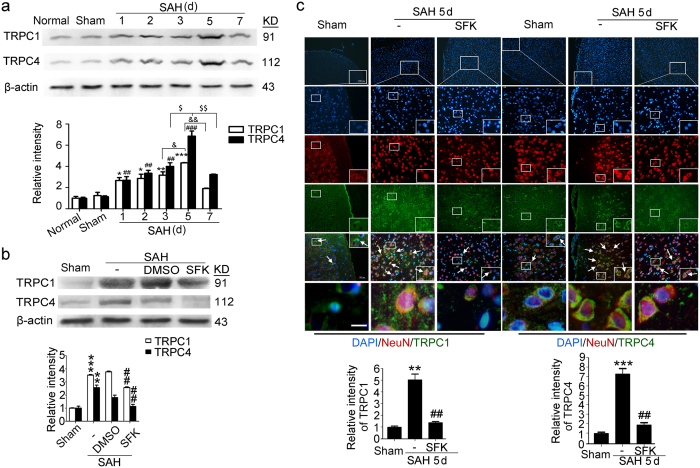
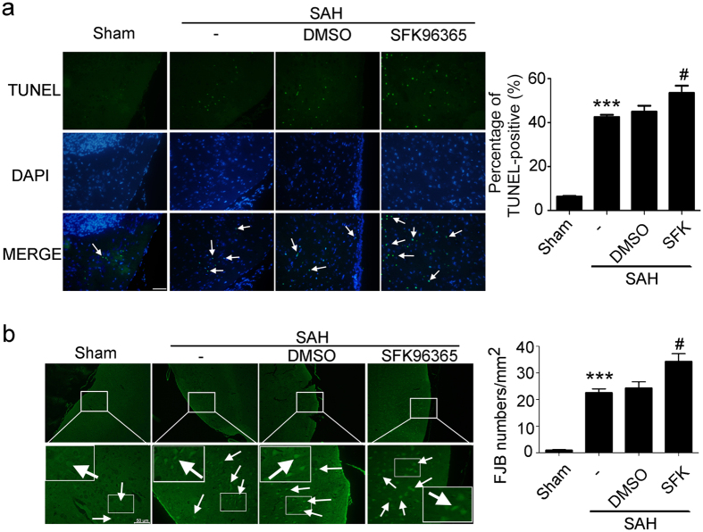
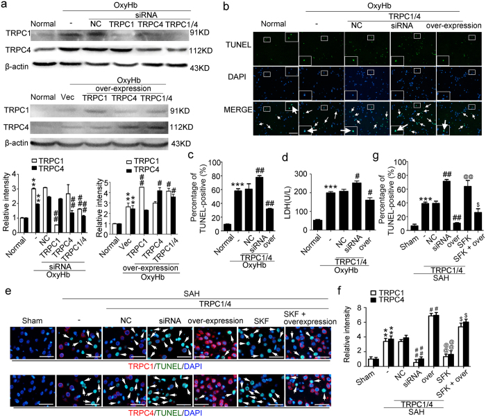
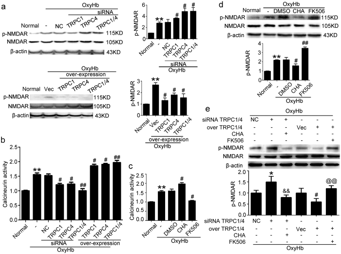
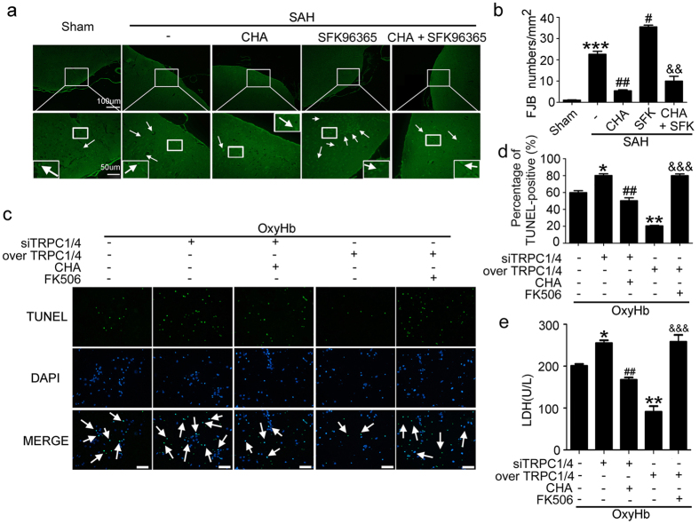
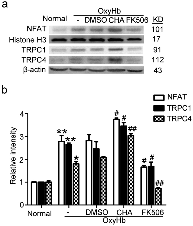
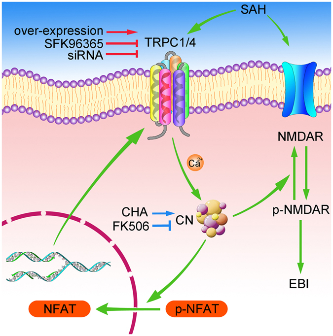
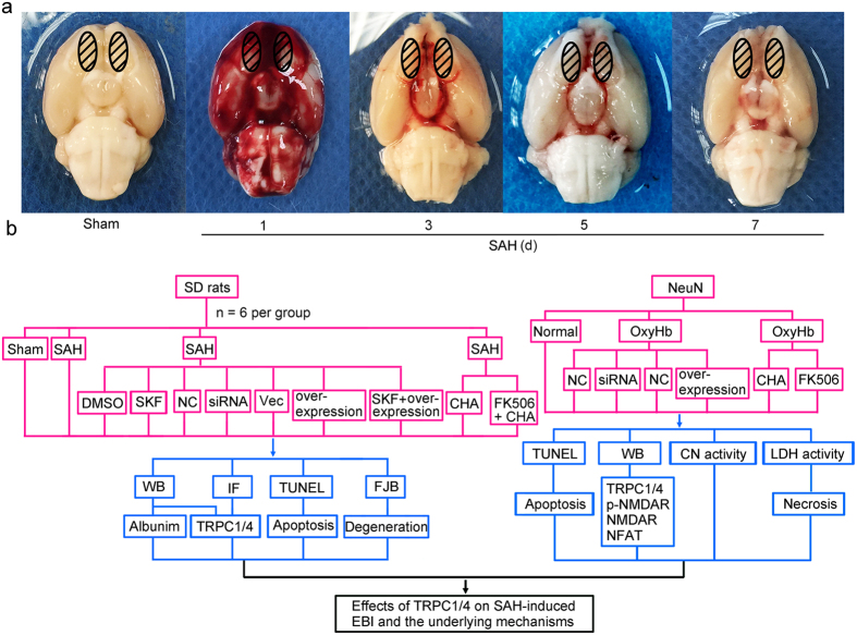
Transient receptor potential channel 1/4 (TRPC1/4) are considered to be related to subarachnoid hemorrhage (SAH)-induced cerebral vasospasm. In this study, a SAH rat model was employed to study the roles of TRPC1/4 in the early brain injury (EBI) after SAH. Primary cultured hippocampal neurons were exposed to oxyhemoglobin to mimic SAH in vitro. The protein levels of TRPC1/4 increased and peaked at 5 days after SAH in rats. Inhibition of TRPC1/4 by SKF96365 aggravated SAH-induced EBI, such as cortical cell death (by TUNEL staining) and degenerating (by FJB staining). In addition, TRPC1/4 overexpression could increase calcineurin activity, while increased calcineurin activity could promote the dephosphorylation of N-methyl-D-aspartate receptor (NMDAR). Calcineurin antagonist FK506 could weaken the neuroprotection and the dephosphorylation of NMDAR induced by TRPC1/4 overexpression. Contrarily, calcineurin agonist chlorogenic acid inhibited SAH-induced EBI, even when siRNA intervention of TRPC1/4 was performed. Moreover, calcineurin also could lead to the nuclear transfer of nuclear factor of activated T cells (NFAT), which is a transcription factor promoting the expressions of TRPC1/4. TRPC1/4 could inhibit SAH-induced EBI by supressing the phosphorylation of NMDAR via calcineurin. TRPC1/4-induced calcineurin activation also could promote the nuclear transfer of NFAT, suggesting a positive feedback regulation of TRPC1/4 expressions.


Figures

Figure 1
Figure 1. Effects of subarachnoid hemorrhage (SAH) stimulus and SFK96365 treatment on the protein levels of TRPC1/4.
(a) Western blot assay of the effect of SAH stimulus. Data are means ± SEM. *p < 0.05, **p = 0.0054, ***p < 0.001, ##p < 0.01, ###p < 0.001 vs. sham group; &p = 0.041, &&p = 0.0067, $p = 0.031, $$p = 0.0064 (n = 6). (b) Western blot assay of the effect of SFK96365 treatment. Data are means ± SEM. **p = 0.006, ***p < 0.001 vs. sham group; ##p < 0.01 vs. SAH + DMSO group (n = 6). (c) Double-immunofluorescence. TRPC1 or TRPC4 (green) and neuronal marker (NeuN, red), and nuclei were fluorescently labeled with DAPI (blue). Arrows point to TRPC1-positive or TRPC4-positive neuronal cells. Scan bar = 32 μm. The relative fluorescent intensity of TRPC1 or TRPC4 in neuronal cells was shown. Data are means ± SEM. In TRPC1, **p = 0.003 vs. sham group, ##p = 0.004 vs. SAH group; In TRPC4,***p < 0.001 vs. sham group, ##p = 0.0051 vs. SAH group, n = 6.
Figure 2
Figure 2. Effects of TRPC1/4 on early brain injury induced by subarachnoid hemorrhage (SAH).
(a) Double staining of TUNEL (green) and DAPI (blue). Arrows point to TUNEL-positive cells in the brain. Scan bar = 100 μm. Data are means ± SEM. ***p < 0.001 vs. sham group, #p = 0.049 vs. SAH + DMSO group (n = 6). (b) Fluoro-jade B (FJB) staining. Arrows point to fluoro-jade B-positive cells. Scan bar = 50 μm. Data are means ± SEM. ***p < 0.001 vs. sham group, #p = 0.035 vs. SAH + DMSO group (n = 6).
Figure 3
Figure 3. Effects of TRPC1/4 on cell death following subarachnoid hemorrhage (SAH).
(a) Western blot analysis. Relative protein level was calculated based on densitometry analysis. Data are means ± SEM. **p < 0.01 vs. normal group; ##p < 0.01, #p < 0.05 vs. oxyHb + negative control siRNA (NC) group or oxyHb + vector (vec) group (n = 3). (b,c) Double staining of TUNEL(green) and DAPI (blue) in cultured neurons. Arrows point to TUNEL-positive cells. Scan bar = 100 μm. Data are means ± SEM. ***p < 0.001 vs. normal group; ##p < 0.01 vs. oxyHb + negative control siRNA (NC) group (n = 3). (d) LDH activity in the culture medium. Data are means ± SEM. ***p < 0.001 vs. normal group; #p < 0.05 vs. oxyHb + negative control siRNA (NC) group (n = 3). (e) Combination of TUNEL assay with immunofluorescence assay. Immunostaining of TRPC1 or TRPC4 (red) co-stained with TUNEL (green) in the brain at 72 h after SAH onsets. The nuclei were counterstained with DAPI (blue). Arrows point to TUNEL-positive cells co-labeled with low fluorescence intensity of TRPC1/4. Bar = 60 μm. The relative fluorescent intensity of TRPC1 or TRPC4 was shown in (f). Percentage of TUNEL-positive cells in the brain was shown in (g). Data are means ± SEM. ***p < 0.001, **p < 0.01, #p < 0.05, ##p < 0.01 vs. SAH + negative control siRNA (NC) group, @@p < 0.01 vs. SAH group, $p < 0.05 vs. SAH + SFK 96365 group (n = 6).
Figure 4
Figure 4. TRPC1/4 supressed the phosphorylation of NMDAR via calcineurin in neurons exposed to oxyHb.
(a) Western blot analysis of the phosphorylation level of NMDAR in neurons. Data are means ± SEM. **p < 0.01 vs. normal group; #p < 0.05 vs. oxyHb + negative control siRNA (NC) group or oxyHb + vector (Vec) group (n = 3). (b,c) Relative calcineurin activity. Data are means ± SEM. In (b), **p = 0.0084 vs. normal group; #p < 0.05, ##p < 0.01 vs. oxyHb + negative control (NC) group (n = 3). In (c), **p = 0.0085 vs. normal group; #p < 0.05 vs. oxyHb + DMSO group (n = 3). (d) Western blot analysis of the phosphorylation level of NMDAR in neurons. The mean values of normal groups were normalized to 1.0. Data are means ± SEM. **p = 0.0061 vs. normal group; #p = 0.031, ##p = 0.0085 vs. oxyHb + DMSO group (n = 3). (e) Western blot analysis of the phosphorylation level of NMDAR in neurons. Data are means  ± SEM. *p = 0.039 vs. oxyHb + negative control siRNA (NC) group; &&p = 0.0081 vs. oxyHb + TRPC1/4 siRNA group; #p = 0.029 vs. oxyHb + vector (Vec) group; @@p = 0.0069 vs. oxyHb + TRPC1/4 overexpression group (n = 3). CHA: chlorogenic acid.
Figure 5
Figure 5. Roles of calcineurin in the neuroprotective effects of TRPC1/4 following subarachnoid hemorrhage (SAH).
(a) Fluoro-jade B (FJB) staining. Arrows point to fluoro-jade B-positive cells. (b) Data are means ± SEM. ***p < 0.001 vs. sham group; ##p = 0.0043, #p = 0.033 vs. SAH group; &&p = 0.0051 vs. SAH + SFK group (n = 6). (c) Double staining of TUNEL (green) and DAPI (blue) in cultured neurons. Scan bar = 100 μm. Arrows point to TUNEL-positive cells. (d) Cell death rate. Data are means ± SEM. **p = 0.0051, *p = 0.047 vs. oxyHb group; ##p = 0.0071 vs. oxyHb + TRPC1/4 siRNA group; &&&p < 0.001 vs. oxyHb + TRPC1/4 overexpression group (n = 3). (e) LDH activity in the culture medium. Data are means ± SEM. **p = 0.0068, *p = 0.048 vs. oxyHb group; ##p = 0.0091 vs. oxyHb + TRPC1/4 siRNA group; &&&p < 0.001 vs. oxyHb + TRPC1/4 overexpression group (n = 3). CHA: chlorogenic acid.
Figure 6
Figure 6. Roles of calcineurin in nuclear transfer of NFAT in neurons exposed to oxyHb.
(a,b) Western blot analysis of the protein level of NFAT in nuclear protein and the protein levels of TRPC1/4 in total protein. Data are means ± SEM. **p < 0.01, *p = 0.027 vs. normal group; ##p < 0.01, #p < 0.05 vs. oxyHb + DMSO group (n = 3). CHA: chlorogenic acid.
Figure 7
Figure 7. Mechanism of the protective effect induced by TRPC1 and TRPC4 after SAH.
Following SAH, the expression of TRPC1/4 was increased, which in turn upregulated the activity of calcineurin (CN). CN promoted the dephosphorylation of NMDAR, which can restrict the early brain injury, including cortical death and degeneration due to SAH. With overexpression of TRPC1/4, more apparent protective effects were shown. With inhibition of TRPC1/4 (by inhibitor in vivo or by siRNA in vitro), EBI were aggravated. Activated CN promotes the nuclear transfer of NFAT, which in turn upregulates the expression of TRPC1/4. CHA: chlorogenic acid.
Figure 8
Figure 8. Experimental design.
(a) The representative areas taken for assay. (b) Experiment design. NC: negative control siRNA; Vec: vector; CHA: chlorogenic acid.

Similar articles

Cited by

References

    1. de Rooij N. K., Rinkel G. J., Dankbaar J. W. & Frijns C. J. Delayed cerebral ischemia after subarachnoid hemorrhage: a systematic review of clinical, laboratory, and radiological predictors. Stroke 44, 43–54, doi: 10.1161/STROKEAHA.112.674291 (2013). - DOI - PubMed
    1. Cheng C. et al.. Effect of APOE Gene Polymorphism on Early Cerebral Perfusion After Aneurysmal Subarachnoid Hemorrhage. Transl Stroke Res 6, 446–450, doi: 10.1007/s12975-015-0426-6 (2015). - DOI - PubMed
    1. Song J. et al.. Correlating Cerebral (18)FDG PET-CT Patterns with Histological Analysis During Early Brain Injury in a Rat Subarachnoid Hemorrhage Model. Transl Stroke Res 6, 290–295, doi: 10.1007/s12975-015-0396-8 (2015). - DOI - PubMed
    1. Suzuki H. What is early brain injury? Transl Stroke Res 6, 1–3, doi: 10.1007/s12975-014-0380-8 (2015). - DOI - PubMed
    1. Tewari A., Mahendru V., Sinha A. & Bilotta F. Antioxidants: The new frontier for translational research in cerebroprotection. J Anaesthesiol Clin Pharmacol 30, 160–171, doi: 10.4103/0970-9185.130001 (2014). - DOI - PMC - PubMed

Publication types

MeSH terms

LinkOut - more resources