. 2016 Sep 19:7:12875.
doi: 10.1038/ncomms12875.

Dysferlin-mediated phosphatidylserine sorting engages macrophages in sarcolemma repair

Affiliations

Dysferlin-mediated phosphatidylserine sorting engages macrophages in sarcolemma repair

Volker Middel et al. Nat Commun. .

Abstract

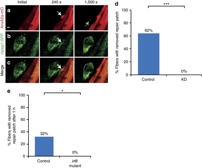
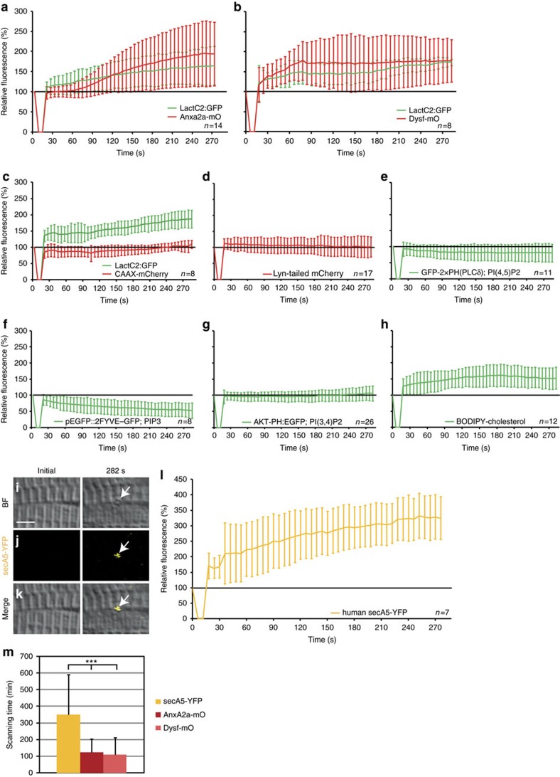
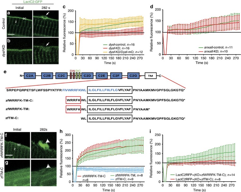
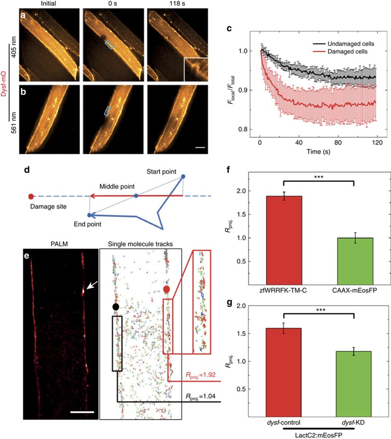
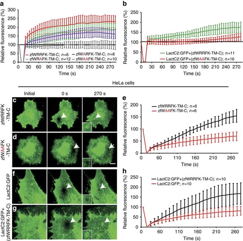
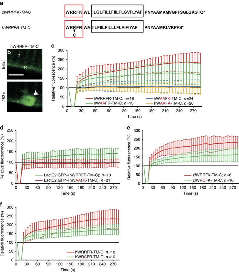
Failure to repair the sarcolemma leads to muscle cell death, depletion of stem cells and myopathy. Hence, membrane lesions are instantly sealed by a repair patch consisting of lipids and proteins. It has remained elusive how this patch is removed to restore cell membrane integrity. Here we examine sarcolemmal repair in live zebrafish embryos by real-time imaging. Macrophages remove the patch. Phosphatidylserine (PS), an 'eat-me' signal for macrophages, is rapidly sorted from adjacent sarcolemma to the repair patch in a Dysferlin (Dysf) dependent process in zebrafish and human cells. A previously unrecognized arginine-rich motif in Dysf is crucial for PS accumulation. It carries mutations in patients presenting with limb-girdle muscular dystrophy 2B. This underscores the relevance of this sequence and uncovers a novel pathophysiological mechanism underlying this class of myopathies. Our data show that membrane repair is a multi-tiered process involving immediate, cell-intrinsic mechanisms as well as myofiber/macrophage interactions.


Figures

Figure 1
Figure 1. Macrophages remove repair patch.
(ac) Repair patch (AnxA2a-mO, a, white arrow) and macrophages (mpeg1:GFP, b, white arrows) before (Initial), 240 s and 1,500 s after wounding. A macrophage ingests parts of repair patch (green arrows). (c) Merged views of a,b. Note the bleed-through of AnxA2a-mO into the GFP channel (b). (d) Repair patch removal in control (left, n=37) and macrophage-depleted embryos (KD, right, n=25, Fisher's exact test P<10−8) over 20 h. (e) Crispr/Cas9 knock-out of irf8 impairs repair patch removal. In controls, the repair patch was removed in 32% of injured myofibers (n=19) within one hour after injury. In the irf8-KO embryos the patch was present in all cases of injured myofibers examined (n=15). This suggests that morpholino triple knock-down is inefficient and the same effect on macrophages can be achieved by elimination of irf8 at the stage analysed. Significance was checked with Fishers exact test at P<0.05 (P=0.023895). Scale bar, 4 μm.
Figure 2
Figure 2. Phosphatidylserine (PS) is sorted to the repair patch.
(ah) Real-time analysis of enrichment of PS (LactC2:GFP (ac)), PI(4,5)P2 (GFP-2 × PH(PLCδ) (e)), PIP3 (pEGFP::2FYVE–GFP (f)), PI(3,4)P2 (AKT-PH:EGFP (g)) and cholesterol (BODIPY-cholesterol (h)), relative to AnxA2a-mO (a), Dysf–mO (b) and membrane markers CAAX-mCherry (c), Lyn-tailed mCherry (d). The reporter fluorescence is expressed as percentage (mean±s.d.) relative to the level before injury. (im) PS is presented on the extracellular side of the repair patch. (il) Extracellularly supplied secA5-YFP was enriched at membrane on lesioning (ik, arrows). secA5-YFP expressing myofibers are outside of the field of view. (l) Kinetics of secA5-YFP at the repair patch. (m) Macrophage scanning-time (+/− s.d.) of myofibers expressing secreted secA5-YFP (yellow, n=19), AnxA2a-mO (dark red, n=22) or Dysf–mO (light red, n=23; Student t-test P<0.001) Scale bar, 4 μm.
Figure 3
Figure 3. Five amino acid motif in Dysf is required for PS accumulation.
(a,b) Before wounding, LactC2:GFP (green) localized to the sarcolemma (sc; a) and the Z-line (z; a) both in control (a) and knock-down (dysf-KD; b) myofibers. On injury, LactC2:GFP accumulated in controls (a, arrow) but not in dysf-KD myofibers (b, arrow). (c) Kinetics of LactC2:GFP accumulation in control-KD (green), dysf-KD (red) and dysf-KD embryos co-injected with mOrange1-DysfC (Dysf–mO, yellow). Lack of PS accumulation in dysf-KD myofibers was rescued by Dysf–mO, translation of which is not inhibited by dysf morpholino. (d) Knock-down of anxa6 (anxa6-KD) had no effect on PS accumulation (red) compared with controls (anxa6-control, green). (e) Domain structure of Dysf. (blue AA: predicted amphipathic helix). (f,g), On damage zfWRRFK-TM-C accumulates in the repair patch (f, arrow). No accumulation was observed for zfTM-C (g, arrow). (h) Accumulation kinetics of zfWRRFK-TM-C (red), zfWRRFK-TM (green) and zfTM-C (blue). A 5-AA motif (WRRFK, red box, e) is required for accumulation. (i) zfWRRFK-TM-C (red) but not zfTM-C (green) rescued PS accumulation in dysf-KD myofibers. In all charts, the change of fluorescence at the lesion is indicated as percentage relative to the undamaged state±s.d.). Scale bars, 4 μm (a,gh), 3 μm (b).
Figure 4
Figure 4. Dysf facilitates PS translocation to site of lesion.
(ac) Fluorescence loss in photobleaching (FLIP) analysis on selected regions (blue boxes in a,b) of live Dysf–mO expressing myofibers. (a) Photobleaching and membrane damaging using 405 nm laser; (b) photobleaching only using 561 nm laser. (c) 561-nm irradiation results in an intensity decrease by 6% (black curve); 405-nm irradiation results in a more rapid decrease by 14% (red curve). (dg) Single-molecule trajectory analysis of zfWRRFK-TM-C and controls after sarcolemmal damage. (d) Scheme showing the projection of a single-molecule trajectory onto the line connecting the midpoint with the site of damage. (e) Super-resolution localization image of a mEosFP:zfWRRFK-TM-C labelled myofiber. The arrow indicates the site of lesion. Single-molecules trajectories calculated from the image data in e. Molecules near the lesion (red box) show a high tendency (Rproj.=1.92) to move towards the lesion (red dot). In the undamaged sarcolemma (left), trajectories (black box) did not show directed motion towards the black dot (Rproj.=1.04). (f) mEosFP:zfWRRFK-TM-C moved towards the lesion (red column); the control CAAX-mEosFP did not (green column; P<10−8). (g) The PS sensor LactC2:mEosFP moved towards the lesion in dysf-contr-KD embryos (red column) but not in dysf-KD embryos (green column; Student t-test P<10−8). n⩾8. Scale bar, 5 μm.
Figure 5
Figure 5. The WRRFK-motif is required for accumulation of Dysf and PS.
(a) After membrane damage, zfWRRFK-TM-C (red) but not zfWAAFK-TM-C (grey) accumulated rapidly at the lesion. The effect was reduced on exchange of the first (zfWARFK-TM-C; purple) or second arginine by alanine (zfWRAFK-TM-C; green). (b) PS (LactC2:GFP) enrichment in the repair patch of Dysf-KD myofibers co-expressing zfWRRFK-TM-C (green), but not of those co-expressing zfWAAFK-TM-C (red). (ce) HeLa cells expressing zfWRRFK-TM-C were imaged before (c) and 0 s (c, arrow) and 270 s after membrane damage (c, arrows). zfWRRFK-TM-C markedly amassed at the lesion (c, arrow, inset; e, black), whereas zfWAAFK-TM-C showed only baseline fluorescence recovery after photobleaching (d, arrows, inset; e, red). (fh) HeLa cells transfected with LactC2:GFP and imaged before (f) and 0 s (arrow) and 270 s after lesioning (arrow) showed no PS accumulation but only baseline recovery (f, arrow, inset; h, red); in the presence of zfWRRFK-TM-C, LactC2:GFP accumulated within the repair patch (g, arrows, inset; h, black). The data in (a,b,e,h) are given as mean±s.e.m. (n=>5) and scaled such that 100% corresponds to fluorescence from the same area before damaging. Scale bars, 10 μm (c,d) and 12.86 μm (f,g).
Figure 6
Figure 6. Arginine-rich motif is mutated in myopathy patients.
(a) Zebrafish zfWRRFK-TM-C and human hWRRFR-TM-C (red box arginine-rich motif; black box: TM domain, arrow: R2042C mutation). (b,c), hWRRFR-TM-C localized to the Z-line (z) and sarcolemma (sc) before damage (b). On damage, it accumulated at the lesion (b, arrow; c, red). In contrast, hWAAFK-TM-C (c, blue) and hWAAFA-TM-C (c, yellow) mutants did not accumulate and hWAAFR-TM-C caused significantly reduced accumulation (c, green). (d) LactC2:RFP accumulated in Dysf-KD myofibers when hWRRFR-TM-C (green) but not when mutant hWAAFK-TM-C (red). was co-expressed. (e,f) zfWRCFK-TM-C (e, green) and hWRCFR-TM-C (f, green), modelling the R2042C dysferlinopathic mutation showed significantly reduced accumulation (red). The fluorescence intensity of Dysf reporters at lesion relative to levels before damage is expressed as mean±s.d. (n⩾6). Note that the control data on hWRRFR-TM-C (c,f) are identical. Similarly, zfWRRFK-TM-C data (e) are also shown in Figs 3h and 5a. Scale bar, 4 μm.

References

    1. Bansal D. et al.. Defective membrane repair in dysferlin-deficient muscular dystrophy. Nature 423, 168–172 (2003). - PubMed
    1. Cooper S. T. & McNeil P. L. Membrane repair: mechanisms and pathophysiology. Physiol. Rev. 95, 1205–1240 (2015). - PMC - PubMed
    1. Bashir R. et al.. A gene related to Caenorhabditis elegans spermatogenesis factor fer-1 is mutated in limb-girdle muscular dystrophy type 2B. Nat. Genet. 20, 37–42 (1998). - PubMed
    1. Liu J. et al.. Dysferlin, a novel skeletal muscle gene, is mutated in Miyoshi myopathy and limb girdle muscular dystrophy. Nat. Genet. 20, 31–36 (1998). - PubMed
    1. Therrien C., Di Fulvio S., Pickles S. & Sinnreich M. Characterization of lipid binding specificities of dysferlin C2 domains reveals novel interactions with phosphoinositides. Biochemistry 48, 2377–2384 (2009). - PubMed

Publication types

Supplementary concepts