. 2017 Feb;21(2):349-363.
doi: 10.1111/jcmm.12969. Epub 2016 Sep 19.

Type 2 diabetes alters mesenchymal stem cell secretome composition and angiogenic properties

Affiliations

Type 2 diabetes alters mesenchymal stem cell secretome composition and angiogenic properties

Jonathan Ribot et al. J Cell Mol Med. 2017 Feb.

Abstract

This study aimed at characterizing the impact of type 2 diabetes mellitus (T2DM) on the bone marrow mesenchymal stem cell (BMMSC) secretome and angiogenic properties. BMMSCs from Zucker diabetic fatty rats (ZDF) (a T2DM model) and Zucker LEAN littermates (control) were cultured. The supernatant conditioned media (CM) from BMMSCs of diabetic and control rats were collected and analysed. Compared to results obtained using CM from LEAN-BMMSCs, the bioactive content of ZDF-BMMSC CM (i) differently affects endothelial cell (HUVEC) functions in vitro by inducing increased (3.5-fold; P < 0.01) formation of tubule-like structures and migration of these cells (3-fold; P < 0.001), as well as promotes improved vascular formation in vivo, and (ii) contains different levels of angiogenic factors (e.g. IGF1) and mediators, such as OSTP, CATD, FMOD LTBP1 and LTBP2, which are involved in angiogenesis and/or extracellular matrix composition. Addition of neutralizing antibodies against IGF-1, LTBP1 or LTBP2 in the CM of BMMSCs from diabetic rats decreased its stimulatory effect on HUVEC migration by approximately 60%, 40% or 40%, respectively. These results demonstrate that BMMSCs from T2DM rats have a unique secretome with distinct angiogenic properties and provide new insights into the role of BMMSCs in aberrant angiogenesis in the diabetic milieu.

Keywords: MSCs; angiogenesis; diabetes type 2; endothelial cells; secretome.


Figures

Figure 1
Figure 1
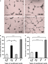
Conditioned supernatant medium from BMMSCs of diabetic rats promotes tubular‐like formation by HUVECs. (A) Representative images of HUVECs in CMLEAN, CMZDF, FM or NCM cultured on Matrigel for 24 hrs showing formation of tubular‐like structures. Scale bar = 100 μm. (B) Quantification of the tubular‐like structures formed by HUVECs in CMLEAN, CMZDF, FM or NCM cultured on Matrigel. Data are expressed as a percentage of the results obtained with CMLEAN (group control). (C) Protein content in the supernatant CMLEAN, CMZDF, FM and NCM. Values are mean ± SEM of four randomly selected fields on each Matrigel surface tested. **P < 0.01, ***P < 0.001. CMLEAN: supernatant, conditioned medium from BMMSCs of control rats. CMZDF: supernatant, conditioned medium from BMMSCs of diabetic rats. FM: Alpha MEM containing 10% foetal bovine serum (FBS) not exposed to cells. NCM: FBS‐free alpha MEM not exposed to cells.
Figure 2
Figure 2
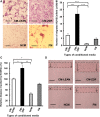
Conditioned supernatant medium from BMMSCs of diabetic rats induces increased HUVEC migration. HUVEC migration was determined using Boyden chambers. (A) Representative images showing HUVEC migration. Stain: Giemsa. Scale bar = 200 μm. (B) Number of migrated HUVECs in the presence of CMLEAN, CMZDF, FM or NCM. HUVECs using the in vitro scratch wound‐healing assay. (C) Distance of migrated HUVECs exposed to supernatant CMLEAN, CMZDF, FM or NCM. (D) Representative light micrographs of the scratch area after 6 hrs. The dotted lines delineate the original scratched area after 6 hrs. Scale bar = 100 μm. Data are expressed as a percentage of the results obtained from the control (CMLEAN) group. Values are mean ± SEM of four fields on each surface tested. *P < 0.05, **P < 0.01, ***P < 0.001. CMLEAN: supernatant, conditioned medium from BMMSCs of control rats. CMZDF: supernatant, conditioned medium from BMMSCs of diabetic rats. FM: alpha MEM containing 10% foetal bovine serum (FBS), not exposed to cells. NCM: FBS‐free alpha MEM not exposed to cells.
Figure 3
Figure 3
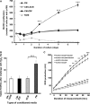
Conditioned supernatant medium from BMMSCs of either diabetic or healthy rats has no effect on either HUVEC proliferation or viability when challenged by H2O2, and a limited effect on reactive oxygen species production. (A) Proliferation of HUVECs in the presence of supernatant CMLEAN, CMZDF, FM and NCM cultured for up to 7 days. (B) HUVEC apoptosis determined using the MTT assay. HUVECs were exposed to 150 μM of H2O2 in supernatant CMLEAN, CMZDF, FM or NCM for 90 min. (C) ROS production by HUVECs exposed to either CMLEAN or CMZDF for 5 hrs. A.U.: arbitrary units. Values are mean ± SEM. ***P < 0.001. CMLEAN: supernatant, conditioned medium from BMMSCs of control rats. CMZDF: supernatant, conditioned medium from BMMSCs of diabetic rats. FM: alpha MEM containing 10% foetal bovine serum (FBS) not exposed to cells. NCM: FBS‐free alpha MEM not exposed to cells.
Figure 4
Figure 4
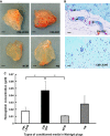
Conditioned supernatant medium from BMMSCs of diabetic rats enhances in vivo angiogenesis. Matrigel plugs (500 μl) containing proteins (500 μg) from freeze‐dried supernatant CMZDF, CMLEAN, FM or NCM were injected subcutaneously into the flanks of nude mice. After 14 days, these Matrigel plugs were excised and photographed, and their haemoglobin contents were quantified. (A) Representative photographs of the excised Matrigel plugs. Scale bar = 1 mm. (B) Representative photograph of sectioned paraffin‐embedded Matrigel plugs stained with Masson's trichrome (the arrowhead indicates capillaries with red blood cells). Scale bar = 20 μm (C) Quantification of the haemoglobin contents in the excised Matrigel plugs. Values are mean ± SEM; n = 6 per experimental group. *P < 0.05. CMLEAN: supernatant, conditioned medium from BMMSCs of control rats. CMZDF: supernatant, conditioned medium from BMMSCs of diabetic rats. FM: alpha MEM containing 10% FBS not exposed to cells. NCM: FBS‐free alpha MEM not exposed to cells.
Figure 5
Figure 5
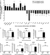
Expression of angiogenic gene mediators is modified in BMMSCs of diabetic rats. (A) Expression of select pro‐angiogenic genes (black bars) and anti‐angiogenic genes (grey bars) in ZDFMSCs relative to results obtained from LEANMSsC. (B) ELISA results of the analysis of overexpressed pro‐angiogenic genes in samples from ZDFMSCs. Values are mean ± SEM. n = 3. ***P < 0.001. LEANMSCs: BMMSCs from control rats. ZDFMSC: BMMSCs from diabetic rats. CMLEAN: supernatant, conditioned medium from BMMSCs of control rats. CMZDF: supernatant conditioned medium from BMMSCs of diabetic rats.
Figure 6
Figure 6
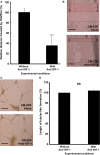
IGF‐1 in CMZDF promotes migration of HUVECs but not tubular formation. HUVEC migration assays were performed using the in vitro scratch/wound‐healing assay. (A) Distance of migrated HUVECs exposed to either supernatant CMZDF or CMZDF containing a blocking antibody for IGF‐1. (B) Representative light micrographs of the scratch area after 6 hrs. The dotted lines delineate the original scratched area after 6 hrs. Scale bar = 100 μm. (C) Representative images of HUVECs in either CMZDF without or CMZDF with a blocking antibody for IGF‐1 (anti‐IGF‐1) cultured on Matrigel for 24 hrs. Scale bar = 100 μm. (D) Quantification of the tubular‐like structures formed by HUVECs in either CMZDF without or CMZDF with blocking antibody for IGF‐1 cultured on Matrigel. Data are expressed as a percentage of the results obtained with CMZDF without anti‐IGF‐1 (control group). Values are mean ± SEM of four fields on each surface tested. ***P < 0.001.
Figure 7
Figure 7
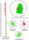
The content of angiogenic mediators in conditioned medium (CM) from BMMSCs of diabetic rats and of controls is different Proteomic analysis of the protein content in supernatant CMZDF compared to the results obtained from CMLEAN. Down‐regulated and overexpressed proteins are shown in red and green, respectively, in all frames. (A) Proteins that were differentially expressed in supernatant CMZDF compared to CMLEAN (B) Proteins present in the supernatant CMZDF that are related to the extracellular matrix. (C) Proteins present in the supernatant CMZDF that are related to glucose metabolism. (D) Proteins present in the supernatant CMZDF that are related to angiogenesis. Values are mean ± SEM. n = 3. P < 0.05. CMLEAN: supernatant, conditioned medium from BMMSCs of control rats. CMZDF: supernatant, conditioned medium from BMMSCs of diabetic rats.
Figure 8
Figure 8
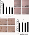
Specific extracellular matrix proteins (LTBP‐1 and LTBP‐2) are involved in the enhanced migration of CMZDF. HUVEC migration assays were performed using the in vitro scratch/wound‐healing assay. (A) Distances migrated by HUVECs exposed to either supernatant CMZDF or CMZDF in the presence or absence of blocking antibodies for either LTBP‐1 or LTBP‐2 or both. (B) Representative light micrographs of the scratch area 6 hrs post cell injury. The dotted lines delineate the original scratched area after 6 hrs. Scale bar = 100 μm. (C) Representative images of HUVECs in either CMZDF or CMZDF and in the presence or absence of blocking antibodies for LTBP‐1, LTBP‐2 or both, cultured on Matrigel for 24 hrs. Scale bar = 100 μm. (D) Quantification of the tubular‐like structures formed by HUVECs exposed to either CMZDF or CMZDF in the presence or absence of blocking antibodies for LTBP‐1, LTBP‐2, or both, cultured on Matrigel for 24 hrs. Data are expressed as percentage of the results obtained when the cells were cultured in CMZDF (control group). Values are mean ± SEM of four fields on each surface tested. *P < 0.05, **P < 0.01.

Similar articles

Cited by

References

    1. Martin A, Komada MR, Sane DC. Abnormal angiogenesis in diabetes mellitus. Med Res Rev. 2003; 23: 117–45. - PubMed
    1. Oikawa A, Siragusa M, Quaini F, et al Diabetes mellitus induces bone marrow microangiopathy. Arterioscler Thromb Vasc Biol. 2010; 30: 498–508. - PMC - PubMed
    1. Tahergorabi Z, Khazaei M. Imbalance of angiogenesis in diabetic complications: the mechanisms. Int J Prev Med. 2012; 3: 827–38. - PMC - PubMed
    1. Abu El‐Asrar AM, Nawaz MI, Kangave D, et al Angiogenesis regulatory factors in the vitreous from patients with proliferative diabetic retinopathy. Acta Diabetol. 2013; 50: 545–51. - PubMed
    1. Abu El‐Asrar AM, Struyf S, Kangave D, et al Chemokines in proliferative diabetic retinopathy and proliferative vitreoretinopathy. Eur Cytokine Netw. 2006; 17: 155–65. - PubMed

MeSH terms