. 2016 Sep 19:6:33489.
doi: 10.1038/srep33489.

Complement component 3 deficiency prolongs MHC-II disparate skin allograft survival by increasing the CD4(+) CD25(+) regulatory T cells population

Affiliations

Complement component 3 deficiency prolongs MHC-II disparate skin allograft survival by increasing the CD4(+) CD25(+) regulatory T cells population

Quan-You Zheng et al. Sci Rep. .

Abstract

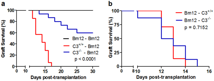
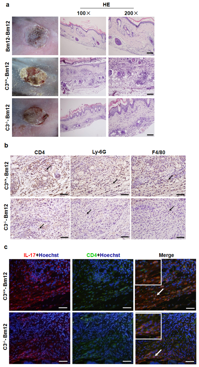
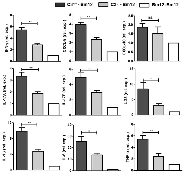
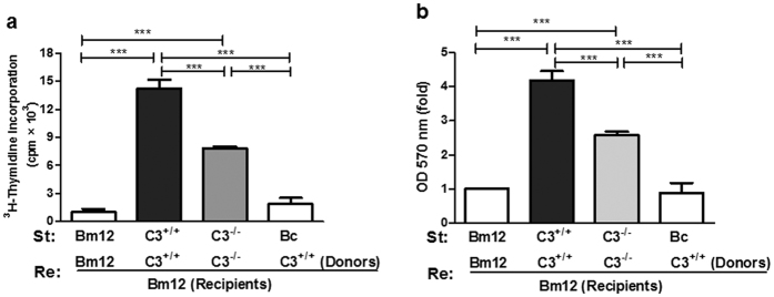
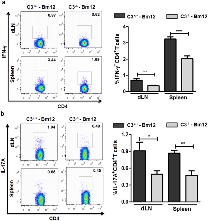
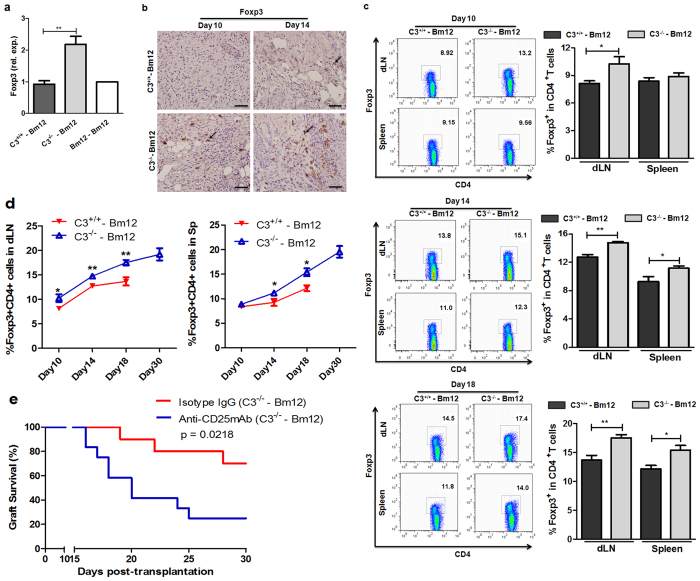
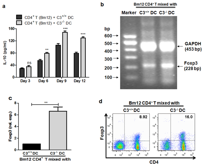
Recent reports suggest that complement system contributes to allograft rejection. However, its underlying mechanism is poorly understood. Herein, we investigate the role of complement component 3 (C3) in a single MHC-II molecule mismatched murine model of allograft rejection using C3 deficient mice (C3(-/-)) as skin graft donors or recipients. Compared with C3(+/+) B6 allografts, C3(-/-) B6 grafts dramatically prolonged survival in MHC-II molecule mismatched H-2(bm12) B6 recipients, indicating that C3 plays a critical role in allograft rejection. Compared with C3(+/+) allografts, both Th17 cell infiltration and Th1/Th17 associated cytokine mRNA levels were clearly reduced in C3(-/-) allografts. Moreover, C3(-/-) allografts caused attenuated Th1/Th17 responses, but increased CD4(+)CD25(+)Foxp3(+) regulatory T (Treg) cell expression markedly in local intragraft and H-2(bm12) recipients. Depletion of Treg cells by anti-CD25 monoclonal antibody (mAb) negated the survival advantages conferred by C3 deficiency. Our results indicate for the first time that C3 deficiency can prolong MHC-II molecule mismatched skin allograft survival, which is further confirmed to be associated with increased CD4(+) CD25(+) Treg cell population expansion and attenuated Th1/Th17 response.


Figures

Figure 1
Figure 1. Local C3 deficiency prolonged MHC-II disparate skin allograft survival.
Mice were transplanted with full thickness tail skin from indicated donors, and transplant rejection was monitored. (a) Alloreactive C3+/+ B6 or C3−/− B6 skin grafts were transplanted into MHC-II disparate Bm12 recipient mice. Syngeneic Bm12 donors were used as negative controls (n = 6–15). (b) Bm12 mice skin grafts were transplanted into C3+/+ or C3−/− B6 recipient mice (n = 7–8). Data were obtained from two independent experiments.
Figure 2
Figure 2. Decreased inflammatory cell infiltration in C3−/− allografts.
Bm12 mice were transplanted with C3+/+, C3−/− or Bm12 grafts. Rejection monitored from day 7 and following experiments performed at day 10 after transplantation. (a) Macroscopic aspects of skin grafts and histology of graft sections stained with haematoxylin and eosin. (b) Skin allografts were stained for CD4, Ly-6G and F4/80. Magnification: ×400. (c) Th17 cell infiltration in allografts was determined by immunofluorescence. Grafts sections were stained with anti-CD4 antibody (green), anti-IL-17 antibody (red), and Hoechst 33258 for nucleus (blue). Magnification: ×400. Clearly positive stained cells were pointed out with arrows (Scale bar: 5 μm).
Figure 3
Figure 3. C3 deficiency led to reduced inflammatory cytokine and chemotactic factor expression in allografts.
Bm12 mice were transplanted with tail skin grafts from Bm12 (Isografts), C3+/+ and C3−/− (allografts) mice. Grafts were harvested on day 10 after transplantation and expression of inflammatory cytokine and chemokine mRNA levels in grafts were measured by RT-qPCR (n = 5–6). One representative data set from three independent experiments is shown. *p < 0.05; **p < 0.01.
Figure 4
Figure 4. C3−/− allografts induced impaired T cell proliferation in Bm12 recipient mice.
Bm12 mice were transplanted with tail skin grafts from Bm12, C3+/+ and C3−/− mice, respectively. Draining lymph node cells (axilla) were collected on day 10 after transplantation and used as the response cells (Re.). The irradiated (2000 rad) naïve splenocytes from Bm12, C3+/+, C3−/− and third-party Bc were used as stimulation cells (St.). The Re. cells were co-cultured with St. cells in 96 well-plates for 72 hours. The cells in culture received 0.5 uCi 3H-thymidine in the last 16 hours and 3H incorporation was determined by liquid scintillation counting (a). T cell proliferation was also measured by MTT assay (b). The data are presented as mean ± SD and are representative of three independent experiments. ***p < 0.001.
Figure 5
Figure 5. C3−/− allograft induced attenuated Th1 and Th17 responses in Bm12 recipient mice.
Bm12 mice were transplanted with tail skin allografts from C3+/+ and C3−/− mice, respectively. Splenocytes and draining lymph node cells (axilla) of Bm12 recipient mice were prepared on day 14 after transplantation, and Th1 (IFN-γ+ CD4+ T) and Th17 (IL-17+ CD4+ T) responses were assessed by FACS. (a) Percentage of IFN-γ+ CD4+ T cells. (b) Percentage of IL-17+ CD4+ T cells. The data are representative of two individual experiments. *p < 0.05; **p < 0.01; ***p < 0.001.
Figure 6
Figure 6. C3−/− allografts led to increased CD4+ CD25+ Treg cell induction in Bm12 recipient mice.
Bm12 mice were transplanted with tail skin grafts from Bm12 (Isografts), C3+/+ and C3−/− (allografts) mice. (a,b) Grafts were harvested at the indicated time points after transplantation. Foxp3 expression in allografts was measured by RT-qPCR on day 10 (a) and IHC on day 10 and day 14 (b). (c,d) Splenocytes and draining lymph node cells (axilla) of Bm12 recipients were prepared at the indicated time points after transplantation and the percentage of Foxp3+ CD4+ T cells was measured by FACS. (e) Neutralizing anti-CD25 mAb or isotype control IgG were administrated to Bm12 recipients with C3−/− tail skin allografts at 0 days, 2 days and 2 weeks post-transplantation. (n = 10–12). The data were representative of three individual experiments. Positive stained cells were pointed out with arrows and the scale bar represents a length of 5 μm. *p < 0.05; **p < 0.01.
Figure 7
Figure 7. C3−/− DCs have increased regulatory T cell driving capacity in vitro.
Following stimulation with LPS for 24 h, the BMDC cells (8 × 104) from either naïve C3+/+ or C3−/− mice were irradiated, and then co-cultured with naïve alloreactive CD4+ T cells (2 × 105) from Bm12 mice for up to 12 days. (a) IL-10 levels in supernatants were examined by ELISA. (b,c) Foxp3 expression was analysed by RT-PCR or qPCR after 9 days co-culture. GAPDH was used as the house keeping gene. The 600-bp DNA markers are shown on the left. (d) The protein levels of Foxp3 in these co-cultured CD4+ T cells were examined by FACS. One representative result from three individual experiments is shown. **p < 0.01; ***p < 0.001.

Similar articles

Cited by

References

    1. Walport M. J. Complement. First of two parts. The New England journal of medicine 344, 1058–1066 (2001). - PubMed
    1. Ricklin D., Hajishengallis G., Yang K. & Lambris J. D. Complement: a key system for immune surveillance and homeostasis. Nature immunology 11, 785–797 (2010). - PMC - PubMed
    1. Kemper C. & Atkinson J. P. T-cell regulation: with complements from innate immunity. Nature reviews. Immunology 7, 9–18 (2007). - PubMed
    1. Carroll M. C. The complement system in regulation of adaptive immunity. Nature immunology 5, 981–986 (2004). - PubMed
    1. Sheen J. H. & Heeger P. S. Effects of complement activation on allograft injury. Current opinion in organ transplantation 20, 468–475 (2015). - PMC - PubMed

Publication types

MeSH terms