

Uroplakin II Expression in Breast Carcinomas Showing Apocrine Differentiation: Putting Some Emphasis on Invasive Pleomorphic Lobular Carcinoma as a Potential Mimic of Urothelial Carcinoma at Metastatic Sites
- PMID: 27642214
- PMCID: PMC5011513
- DOI: 10.1155/2016/2940496
Uroplakin II Expression in Breast Carcinomas Showing Apocrine Differentiation: Putting Some Emphasis on Invasive Pleomorphic Lobular Carcinoma as a Potential Mimic of Urothelial Carcinoma at Metastatic Sites
Abstract
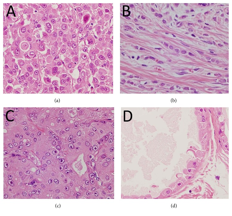
Uroplakin II antibody is exclusively specific for urothelial carcinoma. Nonurothelial carcinoma has not been reported to be immunoreactive for uroplakin II. In the present study, we hypothesized that breast carcinoma showing apocrine differentiation, such as invasive pleomorphic lobular carcinoma (IPLC) and apocrine carcinoma (AC), stains positive for uroplakin II. We identified 6 cases of IPLC between 2000 and 2014 by searching a computerized pathological database. We randomly selected 10 cases of each classic invasive lobular carcinoma (cILC) and AC and five cases of apocrine metaplasia (AM) that coexisted in a surgically resected breast carcinoma specimen. Immunohistochemistry was performed for uroplakin II, GATA3, CK7, CK20, and other representative markers positive for urothelial carcinoma. All cases of IPLC, AC, and AM, except those of cILC, showed immunoreactivity for uroplakin II. Poorly differentiated urothelial carcinoma sometimes shows similar morphology to IPLC with the following immunophenotype: CK7+, CK20-, GATA3+, and uroplakin II+. In the present study, this immunophenotype was observed in all the cases of IPLC and AC. Therefore, when studying metastatic, poorly differentiated carcinoma showing the aforementioned immunophenotype, we should consider the possibility of it being IPLC in addition to metastatic urothelial carcinoma.
Figures

Similar articles
-
Distinct Uroplakin II Staining Pattern in Apocrine Breast Carcinoma.Appl Immunohistochem Mol Morphol. 2022 Nov-Dec 01;30(10):681-686. doi: 10.1097/PAI.0000000000001072. Epub 2022 Oct 14. Appl Immunohistochem Mol Morphol. 2022. PMID: 36227121
-
Uroplakin II (UPII), GATA3, and p40 are Highly Sensitive Markers for the Differential Diagnosis of Invasive Urothelial Carcinoma.Appl Immunohistochem Mol Morphol. 2015 Nov-Dec;23(10):711-6. doi: 10.1097/PAI.0000000000000143. Appl Immunohistochem Mol Morphol. 2015. PMID: 25611245
-
Immunohistochemical Differentiation of Plasmacytoid Urothelial Carcinoma From Secondary Carcinoma Involvement of the Bladder.Am J Surg Pathol. 2017 Nov;41(11):1570-1575. doi: 10.1097/PAS.0000000000000922. Am J Surg Pathol. 2017. PMID: 28786878
-
Apocrine lesions of the breast: part 2 of a two-part review. Invasive apocrine carcinoma, the molecular apocrine signature and utility of immunohistochemistry in the diagnosis of apocrine lesions of the breast.J Clin Pathol. 2019 Jan;72(1):7-11. doi: 10.1136/jclinpath-2018-205485. Epub 2018 Nov 13. J Clin Pathol. 2019. PMID: 30425121 Review.
-
Histiocytoid breast carcinoma: an enigmatic lobular entity.J Clin Pathol. 2011 Aug;64(8):654-9. doi: 10.1136/jcp.2011.088930. Epub 2011 Mar 12. J Clin Pathol. 2011. PMID: 21398688 Review.
Cited by
-
[Mimickers and diagnostic pitfalls of urinary bladder cancer].Pathologie (Heidelb). 2024 Nov;45(6):371-380. doi: 10.1007/s00292-024-01335-4. Epub 2024 May 30. Pathologie (Heidelb). 2024. PMID: 38816588 Review. German.
References
-
- Tian W., Guner G., Miyamoto H., et al. Utility of uroplakin II expression as a marker of urothelial carcinoma. Human Pathology. 2015;46:58–64. - PubMed
-
- Hoang L. L., Tacha D., Bremer R. E., Haas T. S., Cheng L. Uroplakin II (UPII), GATA3, and p40 are highly sensitive markers for the differential diagnosis of invasive urothelial carcinoma. Applied Immunohistochemistry and Molecular Morphology. 2015;23(10):711–716. doi: 10.1097/pai.0000000000000143. - DOI - PubMed
-
- Paner G. P., Annaiah C., Gulmann C., et al. Immunohistochemical evaluation of novel and traditional markers associated with urothelial differentiation in a spectrum of variants of urothelial carcinoma of the urinary bladder. Human Pathology. 2014;45(7):1473–1482. doi: 10.1016/j.humpath.2014.02.024. - DOI - PubMed
MeSH terms
Substances
LinkOut - more resources
Full Text Sources
Other Literature Sources
Medical
Research Materials
