

Isolated localization of Rosai Dorfman disease as renal mass: a case report and review of literature
- PMID: 27642405
- PMCID: PMC5012812
- DOI: 10.11604/pamj.2016.24.64.6291
Isolated localization of Rosai Dorfman disease as renal mass: a case report and review of literature
Abstract
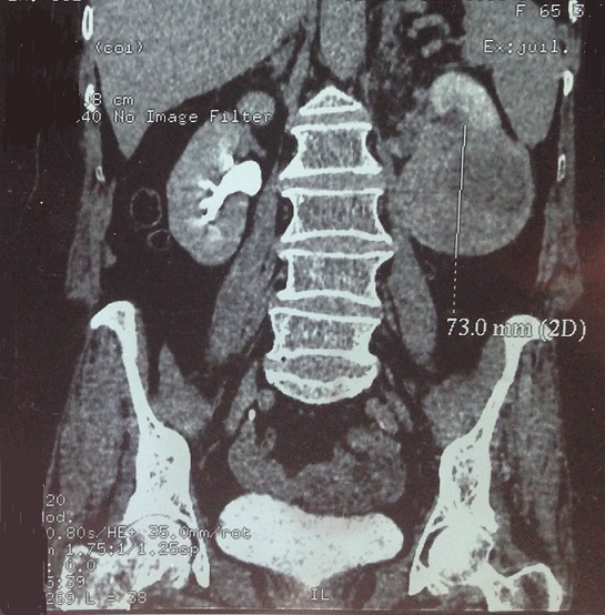
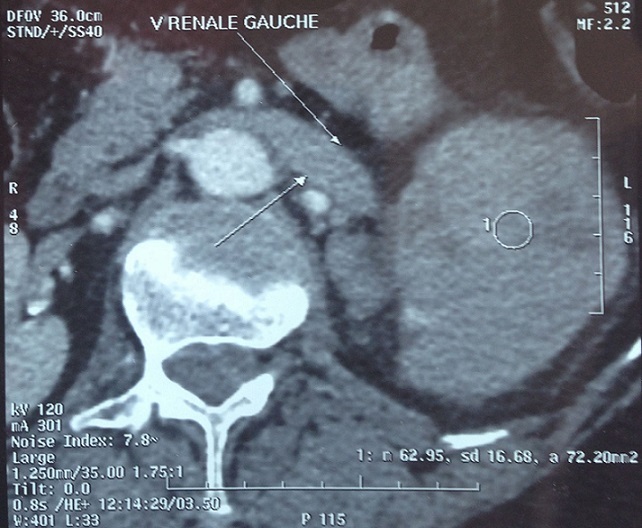
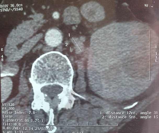
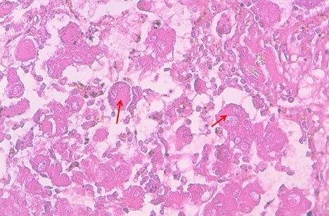
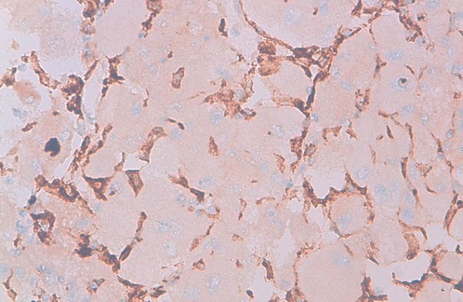
We report a rare case of an elderly woman presented with right renal mass with invasion of renal vein and several small lymphadenopathy in the hilar area. The diagnosis of kidney cancer is suspected and the patient underwent open radical nephrectomy, surrenalectomy and lymphadenectomy dissection. The pathologic examinations find a rosai dorfman disease. This unusual benign entity is uncommon in the kidney, but in medical imaging, it may simulate an infiltrative renal neoplasm, especially a lymphoma or leukemia or even renal cell carcinoma. A comprehensive literature review was undertaken to summarize the clinical and pathologic features of this disorder.
Keywords: Rosai dorfman disease; emperipolesis; extranodal; renal mass.
Figures
References
-
- Rosai J, Dorfman RF. Sinus histiocytosis with massive lymphadenopathy: a newly recognized benign clinicopathologic entity. Arch Path. 1969 Jan;87(1):63–70. - PubMed
-
- Foucar E, Rosai J, Dorfman R. Sinus histiocytosis with massive lymphadenopathy (Rosai-Dorfman disease): review of the entity. Semin Diagn Pathol. 1990 Feb;7(1):19–73. - PubMed
-
- Rosai J, Dorfman RE. Sinus histiocytosis with massive lymphadenopathy: a pseudolymphomatous benign disorder-analysis of 34 cases. Cancer. 1972 Nov;30(5):1174–88. - PubMed
-
- Buchino JJ, Byrd BP, Kmetz DR. Disseminated sinus histiocytosis with massive lymphadenopathy. Arch Pathol Lab Med. 1982 Jan;106(1):13–6. - PubMed
-
- Robert E, Bechtold MD, et al. Renal sinus histiocytosis. Radiol. 1987;162:689–690. - PubMed
Publication types
MeSH terms
LinkOut - more resources
Full Text Sources
Other Literature Sources
Medical
