. 2016 Aug 28:9:100-11.
doi: 10.1016/j.dib.2016.08.046. eCollection 2016 Dec.

Data showing non-conventional HLA-B27 expression in axial joints and gut tissue from B27 transgenic rats, and in frozen and paraffin-fixed synovial SpA tissue

Affiliations

Data showing non-conventional HLA-B27 expression in axial joints and gut tissue from B27 transgenic rats, and in frozen and paraffin-fixed synovial SpA tissue

Oliwia Rysnik et al. Data Brief. .

Abstract

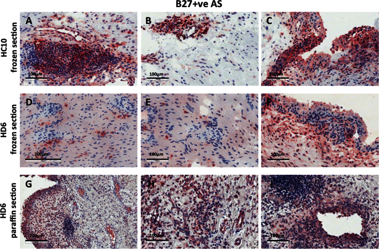
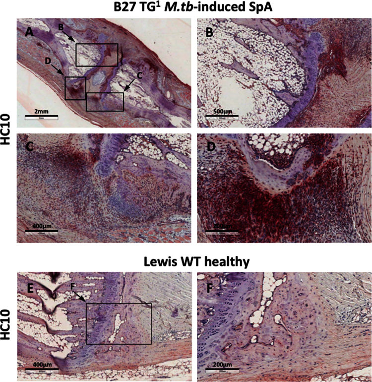
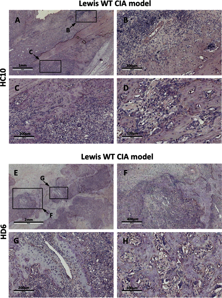
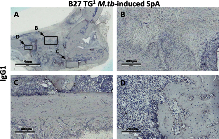
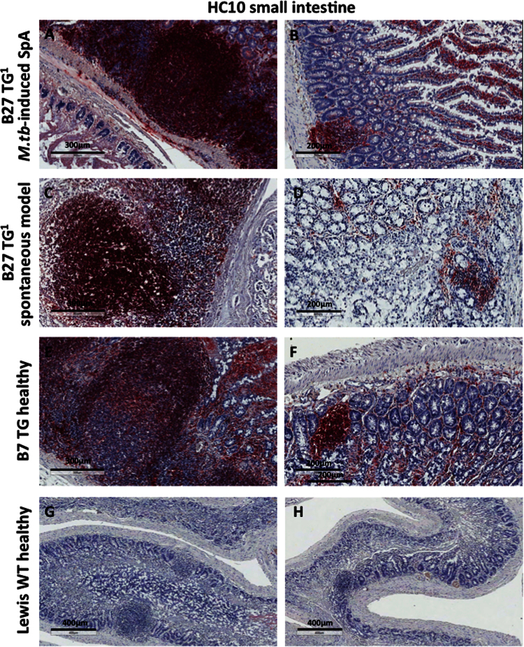
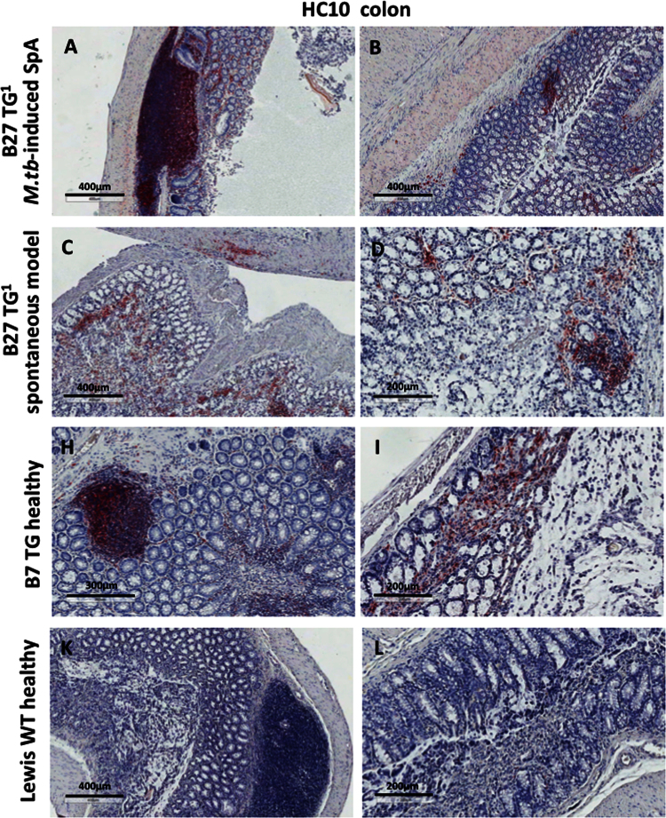
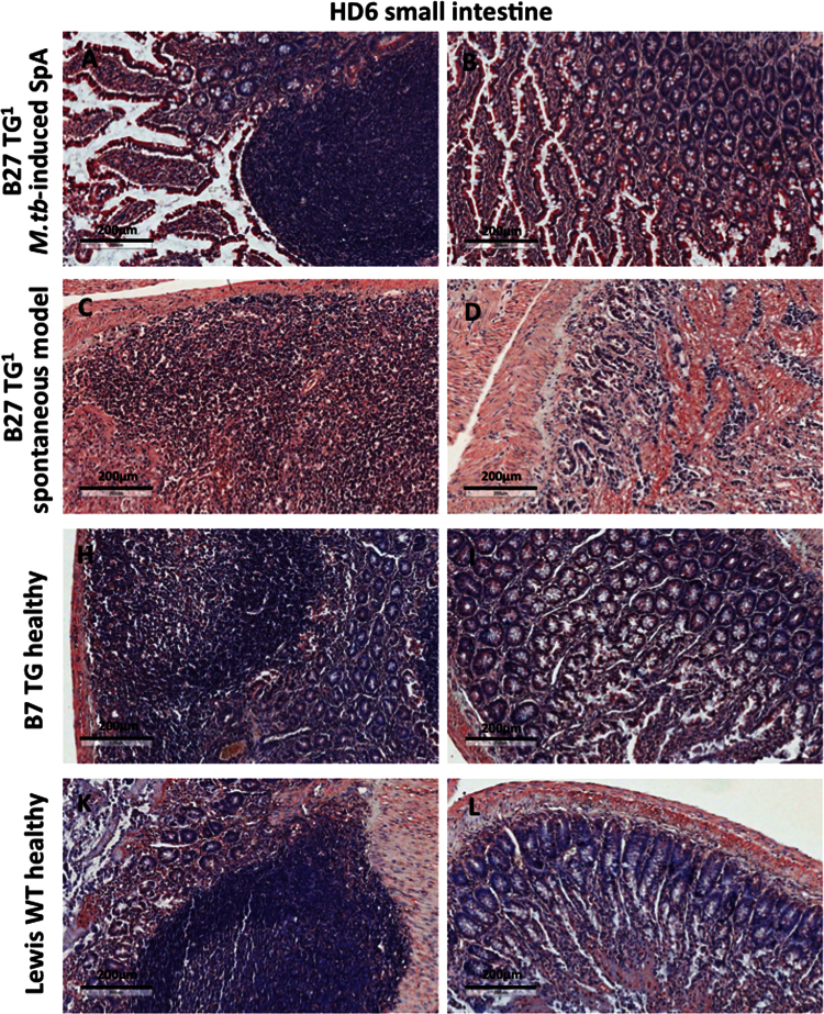
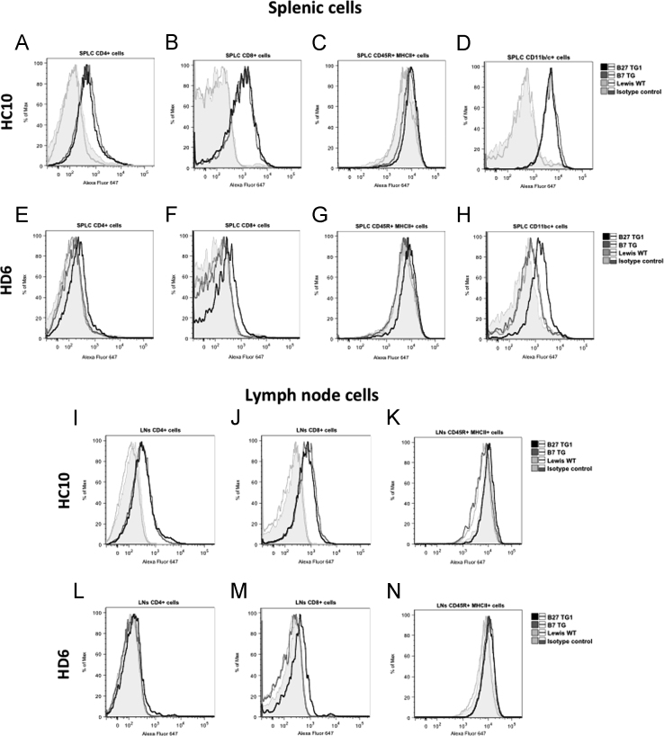
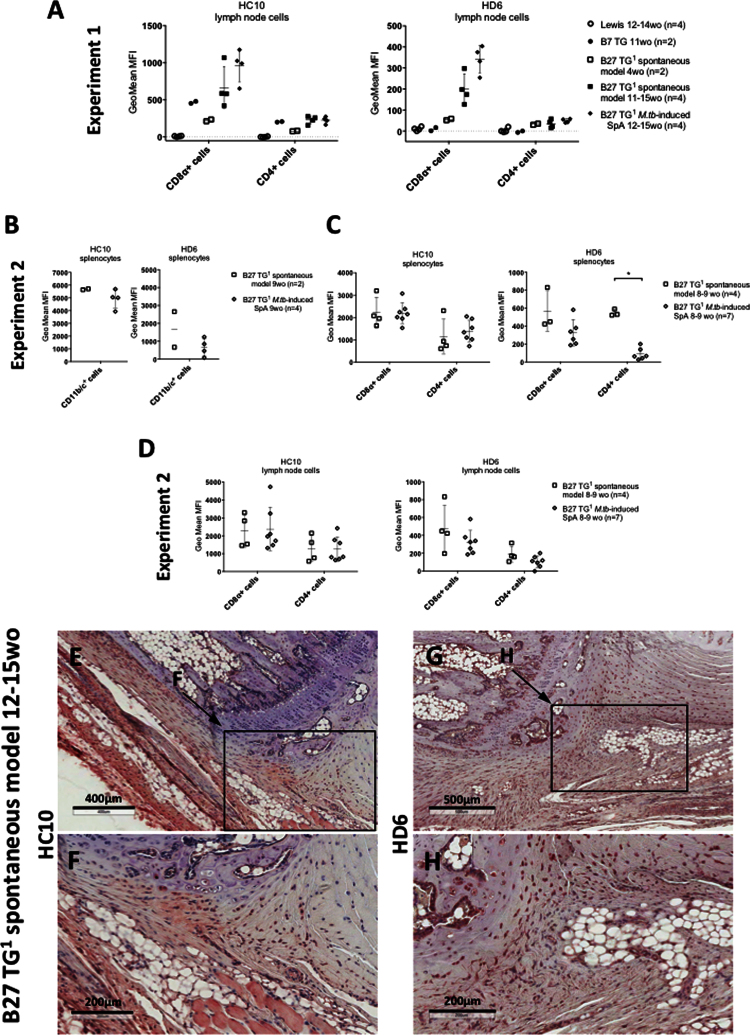
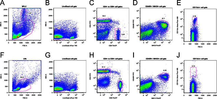
Data is presented showing expression of non-conventional (NC) heavy chain forms of B27 in synovial tissues from SpA patients. Data is presented showing the expression patterns of NC-B27 in joint, gastrointestinal and lymphoid tissues from B27 transgenic (TG(1)) rats with M. tuberculosis-induced SpA. Expression of NC-B27 was determined by immunohistochemistry and flow cytometry using HC10 and HD6 antibodies. These data are the extension of the data presented and discussed in "Non-conventional forms of HLA-B27 are expressed in Spondyloarthritis joints and gut tissue" (O. Rysnik, K. McHugh, L. M. van Duivenvoorde, M. N. van Tok, G. Guggino, J. D. Taurog, S. Kollnberger, F. Ciccia, D. L. Baeten, P. Bowness, 2016) [1].

Keywords: HLA class I free-heavy chains; HLA-B27; HLA-B27 transgenic rat model; Spondyloarthropathies.


Figures

Fig. 1
Fig. 1
shows HC10 and HD6 staining of both frozen and paraffin-fixed synovial tissues from patients with HLA-B27-positive Spondyloarthritis (SpA).
Fig. 2
Fig. 2
(A–D) shows HC10 staining of axial joints from B27 TG1 rats with M.tb-induced SpA. Staining was observed particularly in cell infiltrates at the junction between the vertebrae, connective tissue and annulus fibrosus. We did not observe HC10 staining in ankle or tail joints from healthy Lewis WT rats (Fig. 2E and F).
Fig. 3
Fig. 3
We did not observe HC10 or HD6 staining in ankle joints from Lewis WT rats with adjuvant-induced arthritis (AIA).
Fig. 4
Fig. 4
Shows that tissue sections from B27 TG1 rats with M.tb-induced arthritis and spondylitis did not stain with IgG1 isotype control antibody.
Fig. 5
Fig. 5
Shows HC10 staining was detectable on mononuclear cells in small intestinal Peyer׳s patches, in lymphoid follicles and in the lamina propria of all transgenic animals. Staining levels were higher for B27 TG1 rats with M.tb-induced arthritis and spondylitis compared to those without M.tb or healthy B7 TG animals.
Fig. 6
Fig. 6
Specific HC10 staining was also seen in the colon.
Fig. 7
Fig. 7
HD6 stained small and large bowel tissues, although with background staining observed (Fig. 7 and data not shown). These data show that NC-B27 are expressed in gut tissue in B27 TG1 rats.
Fig. 8
Fig. 8
Shows that splenic and lymph node CD45+/MHCII+ leukocytes from B27 TG1 rats with M.tb-induced arthritis and spondylitis expressed very low levels of NC-B27 molecules.
Fig. 9
Fig. 9
Similar results were observed with cells isolated from B27 TG1 lymph nodes +/− M.tb (Fig. 9A), noting that the CD11b/c+ cell population was absent (see Fig. 10J). We also investigated HC10 and HD6 staining of splenocytes taken from 8–9 weeks old B27 TG1 animals with and without M.tb-induced SpA before the appearance of clinical manifestations. HC10 was not significantly altered in splenic CD11b/c+, CD8α+ or CD4+ cells, or on cell populations from LNs (Fig. 9B–D). However, we observed an increase in HD6 staining on splenic CD4+ cells after M.tb treatment (Fig. 9C right-hand panel). No HC10 or HD6 staining was observed in splenic and LN cells from age-matched Lewis WT rats (data not shown). Splenic and LN cells from age-matched B7 TG rats stained with HC10, but not HD6, to a similar degree compared with B27 TG1 animals (spontaneous model) (Fig. 9A).
Fig. 10
Fig. 10
Shows the gating strategy for analysis of spleen and lymph node cell populations studied by FACS.

Similar articles

Cited by

References

    1. Rysnik O., McHugh K., van Duivenvoorde L.M., van Tok M.N., Guggino G., Taurog J.D., Kollnberger S., Ciccia F., Baeten D.L., Bowness P. Non-conventional forms of HLA-B27 are expressed in Spondyloarthritis joints and gut tissue. J. Autoimmun. 2016 Mar 29. pii: S0896-8411(16)30022-1. - PMC - PubMed
    1. van der Linden S., Valkenburg Ha, Cats a. Evaluation of diagnostic criteria for ankylosing spondylitis. A proposal for modification of the New York criteria. Arthritis Rheumatol. 1984;27(4):361–368. - PubMed
    1. Aletaha D., Neogi T., Silman A.J., Funovits J., Felson D.T., Bingham C.O., Birnbaum N.S., Burmester G.R., Bykerk V.P., Cohen M.D., Combe B., Costenbader K.H., Dougados M., Emery P., Ferraccioli G., Hazes J.M.W., Hobbs K., Huizinga T.W.J., Kavanaugh A., Kay J., Kvien T.K., Laing T., Mease P., Ménard Ha, Moreland L.W., Naden R.L., Pincus T., Smolen J.S., Stanislawska-Biernat E., Symmons D., Tak P.P., Upchurch K.S., Vencovský J.J., Wolfe F., Hawker G. Rheumatoid arthritis classification criteria: an American College of Rheumatology/European League Against Rheumatism collaborative initiative. Arthritis Rheumatol. 2010;62(9):2569–2581. - PubMed
    1. Hammer R.E., Maika S.D., Richardson J.A., Tang J.P., Taurog J.D. Spontaneous inflammatory disease in transgenic rats expressing HLA-B27 and human beta 2m: an animal model of HLA-B27-associated human disorders. Cell. 1990;63:1099–1112. - PubMed
    1. Tran T.M., Dorris M.L., Satumtira N., Richardson J.A., Hammer R.E., Shang J., Taurog J.D. Additional human beta2-microglobulin curbs HLA-B27 misfolding and promotes arthritis and spondylitis without colitis in male HLA-B27-transgenic rats. Arthritis Rheumatol. 2006;54(4):1317–1327. Apr. - PubMed