

Computation of breast ptosis from 3D surface scans of the female torso
- PMID: 27643463
- PMCID: PMC5077640
- DOI: 10.1016/j.compbiomed.2016.09.002
Computation of breast ptosis from 3D surface scans of the female torso
Abstract
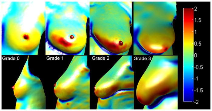
Stereophotography is now finding a niche in clinical breast surgery, and several methods for quantitatively measuring breast morphology from 3D surface images have been developed. Breast ptosis (sagging of the breast), which refers to the extent by which the nipple is lower than the inframammary fold (the contour along which the inferior part of the breast attaches to the chest wall), is an important morphological parameter that is frequently used for assessing the outcome of breast surgery. This study presents a novel algorithm that utilizes three-dimensional (3D) features such as surface curvature and orientation for the assessment of breast ptosis from 3D scans of the female torso. The performance of the computational approach proposed was compared against the consensus of manual ptosis ratings by nine plastic surgeons, and that of current 2D photogrammetric methods. Compared to the 2D methods, the average accuracy for 3D features was ~13% higher, with an increase in precision, recall, and F-score of 37%, 29%, and 33%, respectively. The computational approach proposed provides an improved and unbiased objective method for rating ptosis when compared to qualitative visualization by observers, and distance based 2D photogrammetry approaches.
Keywords: 3D image; Breast surgery; Classification; Gaussian curvature; Histogram matching, Breast ptosis; Orientation; Stereophotogrammetry.
Copyright © 2016 Elsevier Ltd. All rights reserved.
Conflict of interest statement
Conflicts of Interest: None declared.
Figures


Similar articles
-
Assessment of Breast Asymmetry in Adolescent Idiopathic Scoliosis Using an Automated 3D Body Surface Measurement Technique.Spine Deform. 2017 May;5(3):152-158. doi: 10.1016/j.jspd.2017.01.001. Spine Deform. 2017. PMID: 28449957
-
Chances and limitations of a low-cost mobile 3D scanner for breast imaging in comparison to an established 3D photogrammetric system.J Plast Reconstr Aesthet Surg. 2018 Oct;71(10):1417-1423. doi: 10.1016/j.bjps.2018.05.017. Epub 2018 Jun 8. J Plast Reconstr Aesthet Surg. 2018. PMID: 29970344
-
Advancing digital anthropometry in plastic surgery: Comparing smartphone 3D surface imaging to Vectra H2 in breast reconstruction.J Plast Reconstr Aesthet Surg. 2025 May;104:398-406. doi: 10.1016/j.bjps.2025.03.039. Epub 2025 Mar 22. J Plast Reconstr Aesthet Surg. 2025. PMID: 40174257 Clinical Trial.
-
The Three-Dimensional Techniques in the Objective Measurement of Breast Aesthetics.Aesthetic Plast Surg. 2015 Dec;39(6):910-5. doi: 10.1007/s00266-015-0560-2. Epub 2015 Sep 22. Aesthetic Plast Surg. 2015. PMID: 26395095 Review.
-
Use of 3D photography in complex-wound assessment.J Wound Care. 2013 Mar;22(3):156, 158-60. doi: 10.12968/jowc.2013.22.3.156. J Wound Care. 2013. PMID: 23665734 Review.
Cited by
-
Quantitative 3-Dimensional Photographic Assessment of Breast Cosmesis After Whole Breast Irradiation for Early Stage Breast Cancer: A Secondary Analysis of a Randomized Clinical Trial.Adv Radiat Oncol. 2020 May 21;5(5):824-833. doi: 10.1016/j.adro.2020.04.035. eCollection 2020 Sep-Oct. Adv Radiat Oncol. 2020. PMID: 33083644 Free PMC article.
-
An Aesthetic Factor Priority List of the Female Breast in Scandinavian Subjects.Plast Reconstr Surg Glob Open. 2020 Apr 11;8(10):e3173. doi: 10.1097/GOX.0000000000003173. eCollection 2020 Oct. Plast Reconstr Surg Glob Open. 2020. PMID: 33173686 Free PMC article.
-
Three-dimensional breast imaging using Artificial-Intelligence-Based Automatic Measurement System.JPRAS Open. 2025 Feb 3;44:107-118. doi: 10.1016/j.jpra.2025.01.023. eCollection 2025 Jun. JPRAS Open. 2025. PMID: 40160895 Free PMC article.
-
Body image dissatisfaction in patients undergoing breast reconstruction: Examining the roles of breast symmetry and appearance investment.Psychooncology. 2018 Mar;27(3):857-863. doi: 10.1002/pon.4586. Epub 2017 Dec 19. Psychooncology. 2018. PMID: 29152816 Free PMC article.
References
-
- Asgeirsson K, Rasheed T, McCulley S, Macmillan R. Oncological and cosmetic outcomes of oncoplastic breast conserving surgery. Eur J Surg Oncol (EJSO) 2005;31(8):817–823. - PubMed
-
- Grewal NS, Fisher J. Why do patients seek revisionary breast surgery? Aesthet Surg J. 2013;33(2):237–244. - PubMed
-
- Webster R. How does ptosis affect satisfaction after immediate reconstruction plus contralateral mammaplasty? Ann Plast Surg. 2010;65(3):294–299. - PubMed
-
- Fan J, Raposio E, Wang J, Nordström RE. Development of the inframammary fold and ptosis in breast reconstruction with textured tissue expanders. Aesthet Plast Surg. 2002;26(3):219–222. - PubMed
-
- Boutros S, Kattash M, Wienfeld A, Yuksel E, Baer S, Shenaq S. The intradermal anatomy of the inframammary fold. Plast Reconstr Surg. 1998;102(4):1030–1033. - PubMed
MeSH terms
Grants and funding
LinkOut - more resources
Full Text Sources
Other Literature Sources
