. 2016 Nov;186(11):2887-2908.
doi: 10.1016/j.ajpath.2016.07.018.

Accumulation of Ubiquitin and Sequestosome-1 Implicate Protein Damage in Diacetyl-Induced Cytotoxicity

Affiliations

Accumulation of Ubiquitin and Sequestosome-1 Implicate Protein Damage in Diacetyl-Induced Cytotoxicity

Ann F Hubbs et al. Am J Pathol. 2016 Nov.

Abstract

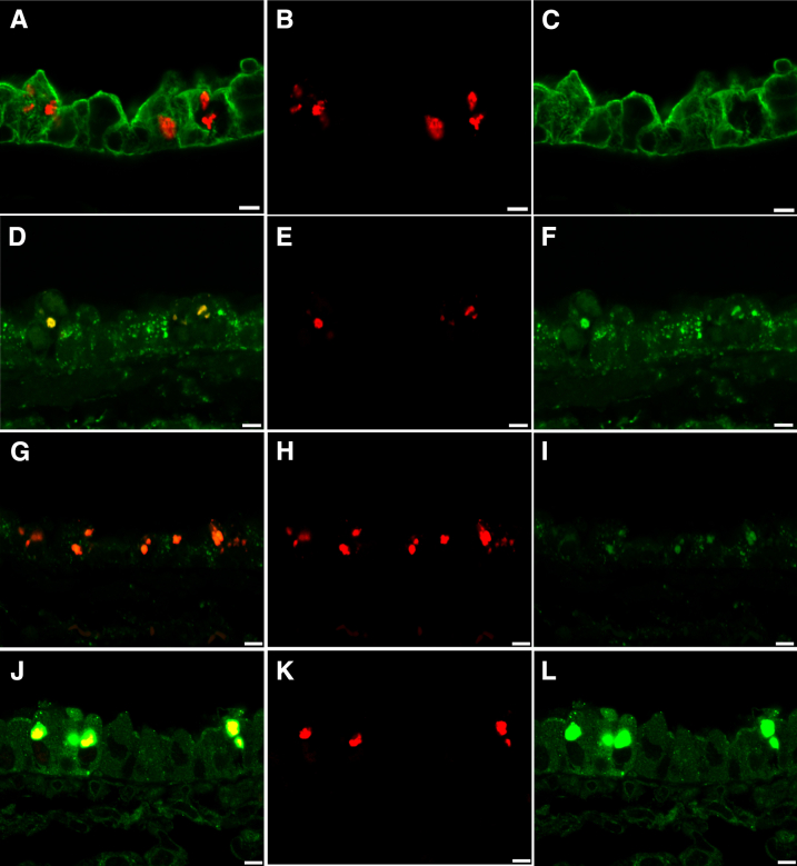
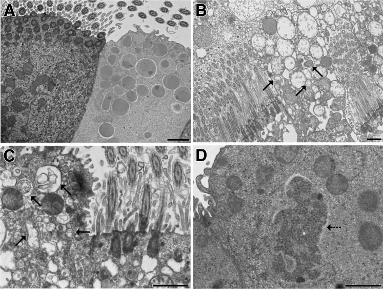
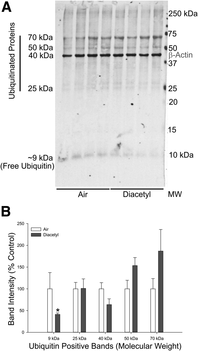
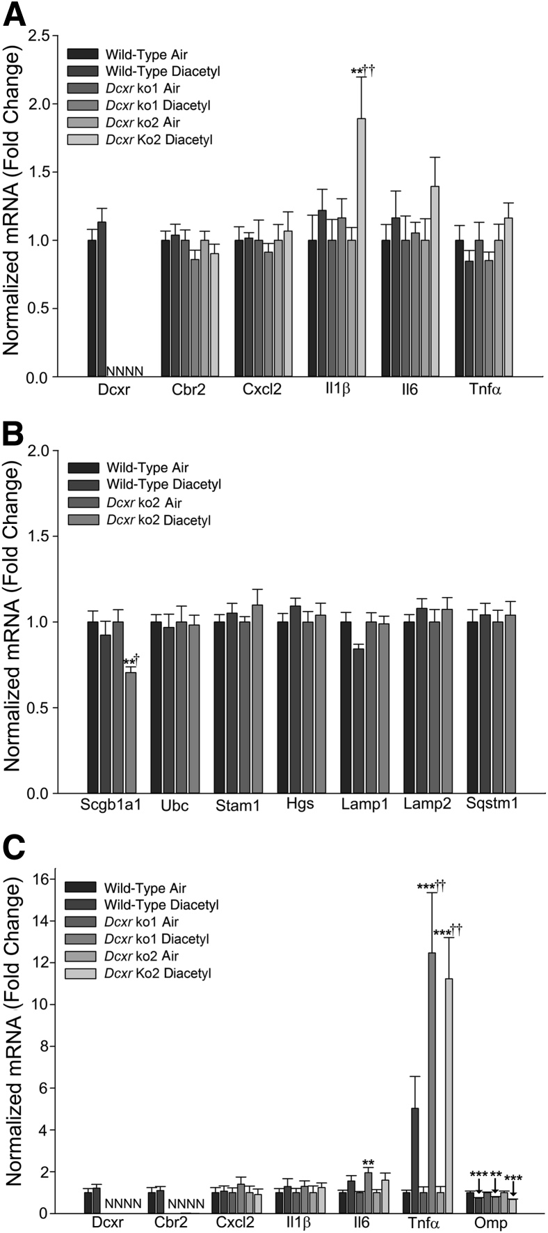
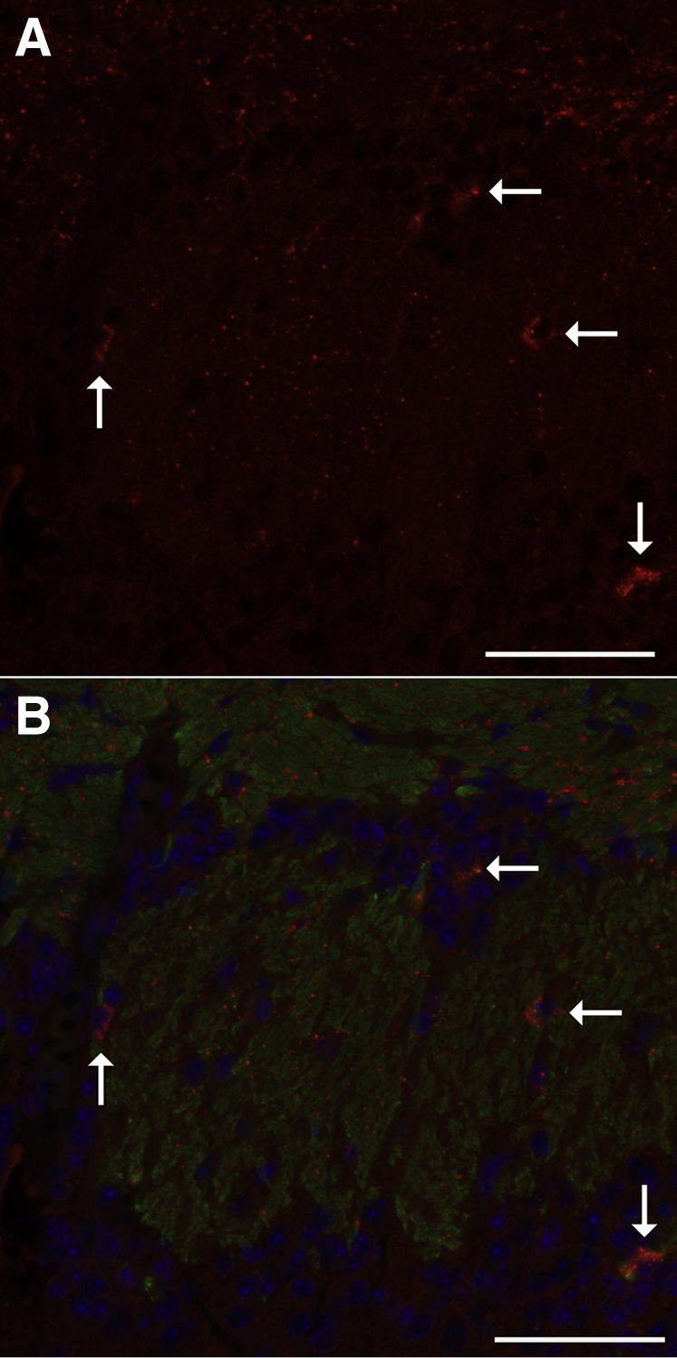
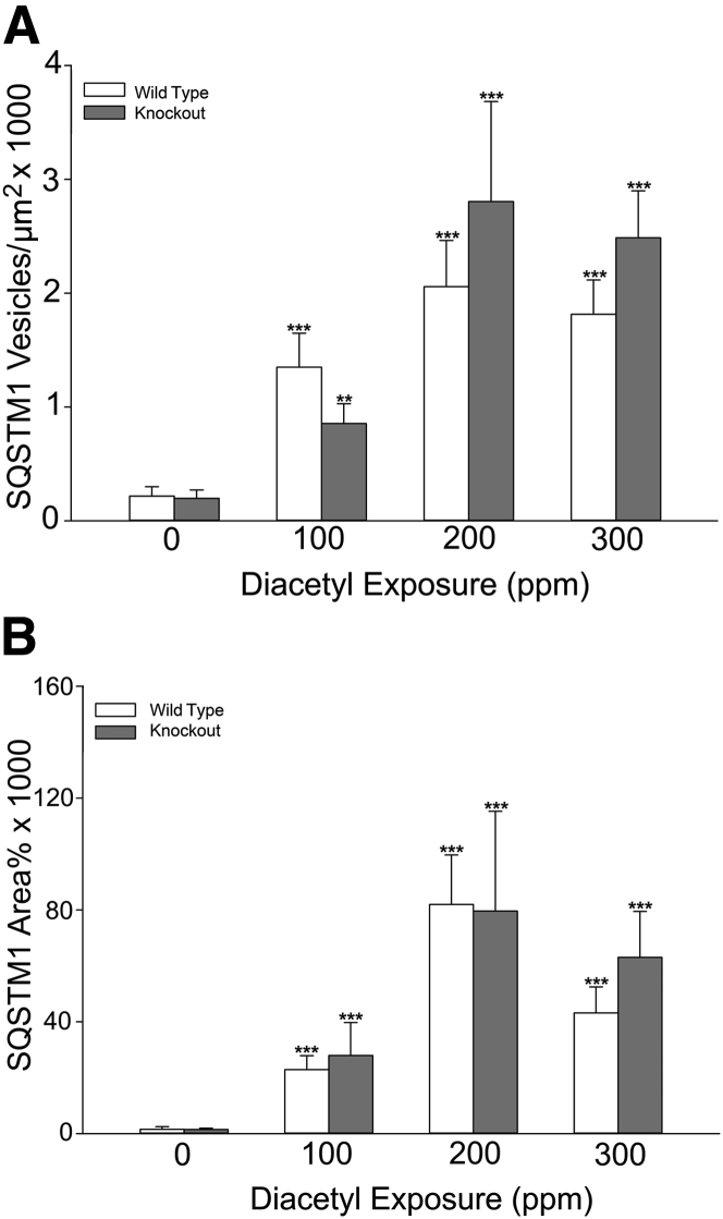
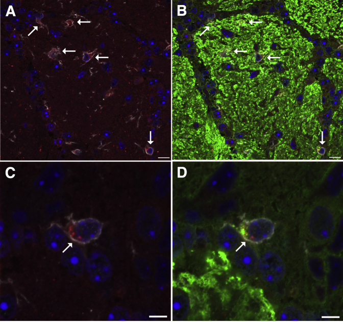
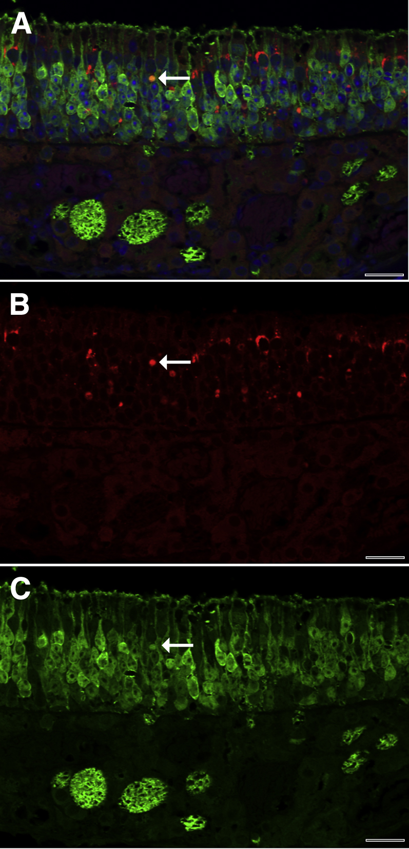
Inhaled diacetyl vapors are associated with flavorings-related lung disease, a potentially fatal airway disease. The reactive α-dicarbonyl group in diacetyl causes protein damage in vitro. Dicarbonyl/l-xylulose reductase (DCXR) metabolizes diacetyl into acetoin, which lacks this α-dicarbonyl group. To investigate the hypothesis that flavorings-related lung disease is caused by in vivo protein damage, we correlated diacetyl-induced airway damage in mice with immunofluorescence for markers of protein turnover and autophagy. Western immunoblots identified shifts in ubiquitin pools. Diacetyl inhalation caused dose-dependent increases in bronchial epithelial cells with puncta of both total ubiquitin and K63-ubiquitin, central mediators of protein turnover. This response was greater in Dcxr-knockout mice than in wild-type controls inhaling 200 ppm diacetyl, further implicating the α-dicarbonyl group in protein damage. Western immunoblots demonstrated decreased free ubiquitin in airway-enriched fractions. Transmission electron microscopy and colocalization of ubiquitin-positive puncta with lysosomal-associated membrane proteins 1 and 2 and with the multifunctional scaffolding protein sequestosome-1 (SQSTM1/p62) confirmed autophagy. Surprisingly, immunoreactive SQSTM1 also accumulated in the olfactory bulb of the brain. Olfactory bulb SQSTM1 often congregated in activated microglial cells that also contained olfactory marker protein, indicating neuronophagia within the olfactory bulb. This suggests the possibility that SQSTM1 or damaged proteins may be transported from the nose to the brain. Together, these findings strongly implicate widespread protein damage in the etiology of flavorings-related lung disease.


Figures

Figure 1
Figure 1
Exposure concentrations as monitored by the volatile organic compound meter. A: Inhalation exposure concentrations for diacetyl during the dose-response study were designed to produce an average chamber concentration of 100 ppm (green), 200 ppm (blue), or 300 ppm (red) of diacetyl. B: Inhalation exposure concentrations for diacetyl during the molecular pathogenesis experiments.
Figure 2
Figure 2
Cellular degeneration and death in the epithelium lining nasal airways and bronchi with corresponding pathology scores. A: Maxilloturbinate from the first nasal section (T1) of a mouse 1 day after inhaling air in an inhalation exposure chamber. The maxilloturbinate is lined by normal transitional epithelium admixed with occasional ciliated epithelial cells. B: Maxilloturbinate from the first nasal section (T1) of a mouse 1 day after inhaling 300 ppm diacetyl. Necrotic cellular debris has replaced the normal transitional epithelium. C: Nasoturbinate from an air-exposed mouse. D: Degenerative changes in respiratory epithelium lining the nasoturbinate of a mouse exposed to 200 ppm diacetyl are characterized by enlarged epithelial cells with rarified cytoplasm and displaced nuclei. E: The pathology score (severity + distribution) for necrosis in the respiratory and transitional epithelium of the nose increased with exposure concentration and decreased as the sections moved away from the front of the nose (T1) sequentially toward the back of the nose (T3). Necrosis was not seen in the air controls (0 ppm). Necrosis was unaffected by sex. Necrosis pathology scores in knockout mice (closed bars) and wild-type mice (open bars) were similar except at 300 ppm at level T1. F: The pathology score (severity + distribution) for necrosis in the olfactory neuroepithelium of the nose increased with diacetyl exposure concentration. Necrosis pathology scores in knockout mice (closed bars) and wild-type mice (open bars) were not significantly different. G: Control bronchus from an air-exposed Dcxr knockout mouse (mouse P11-1126) shows no evidence of degeneration. H: Bronchus of a Dcxr knockout mouse exposed to diacetyl (P11-1129) with epithelial changes characterized by enlargement, cytoplasmic rarefaction, and nuclear displacement. I: Semiquantitative pathology scores from air-exposed control mice demonstrate no evidence of bronchial epithelial degeneration (mean pathology score of 0), whereas diacetyl-exposed mice had bronchial epithelial degeneration, which increased with increasing exposure concentration. Diacetyl significantly increased vacuolar degeneration at all exposure concentrations irrespective of genotype. There was no effect of sex on the diacetyl-induced vacuolar degeneration, and the sexes were combined for analysis of the diacetyl-induced changes. The pathology score for vacuolar degeneration was significantly greater in knockout mice (closed bars) compared to wild-type mice (open bars) at the 200 ppm exposure concentration. J: The number of apoptotic cells detected by the terminal deoxynucleotidyl transferase-mediated dUTP nick-end labeling assay in the epithelium of bronchi was increased in all diacetyl-exposed groups. Apoptosis was not significantly different in knockout (closed bars) compared with wild-type (open bars) mice. P < 0.05, ∗∗P < 0.01, and ∗∗∗P < 0.001 versus air-exposed controls; P < 0.05 versus wild type. Scale bar = 20 μm (AD and GH).
Figure 3
Figure 3
Ubiquitin is increased by diacetyl exposure and localized within intracytoplasmic accumulations in the neuroepithelium of the nose and in intrapulmonary bronchi. A: The olfactory neuroepithelium of a male wild-type mouse exposed to 300 ppm diacetyl, stained by immunofluorescence double labeling for total ubiquitin (red) and OMP (green), demonstrates large ubiquitin (conjugated and unconjugated) accumulations in the neuroepithelium. B: Lung stained by immunofluorescence double label for total ubiquitin (red) and catenin-β1 (green) demonstrates ubiquitin in the airway epithelium of a female wild-type mouse exposed to 300 ppm diacetyl; ubiquitin (conjugated and unconjugated) forms large puncta. C: In the bronchi, diacetyl increased the number of airway epithelial cells containing ubiquitin. The number of cells with ubiquitin puncta was significantly greater in knockout (closed bars) mice compared with wild-type (open bars) mice at the 200 ppm exposure concentration. ∗∗P < 0.01, ∗∗∗P < 0.001 compared with air controls of the same genotype; †††P < 0.001 compared with wild-type mice. Scale bar = 50 μm (A and B).
Figure 4
Figure 4
Dual immunofluorescence for K63-ubiquitin and CDH1. K63-ubiquitin is localized as intraepithelial accumulations in the airway epithelium. The accumulations are associated with diacetyl exposure and are more frequently identified in bronchi than terminal bronchioles. A: Photomicrograph of immunofluorescence double label for K63-ubiquitin (red) and CDH1 (green) in the airway epithelium of the lung of a male Dcxr knockout mouse exposed to 200 ppm diacetyl. K63-ubiquitin forms large puncta similar to those identified by staining for total ubiquitin. B: Morphometric quantification of K63-ubiquitin positive cells in the epithelium of terminal bronchioles of wild-type (open bars) and knockout (closed bars) mice. K63 ubiquitin accumulations were only significantly increased in terminal bronchioles of wild-type mice inhaling 100 ppm. C: Morphometric quantification of K63-ubiquitin positive cells in the epithelium of bronchi. K63-ubiquitin puncta were rare except in diacetyl-exposed mice, and cells containing puncta were significantly increased relative to controls at all exposure concentrations. Knockout mice (closed bars) were significantly more susceptible than wild-type mice (open bars) at 200 ppm when the sexes were pooled for the statistical analysis. P < 0.05, ∗∗∗P < 0.001 compared with air controls of the same genotype; P < 0.05 compared with wild-type mice. Scale bar = 20 μm (A).
Figure 5
Figure 5
Ubiquitin puncta were localized to endosomal/lysosomal/exosomal vesicles and SQSTM1 puncta using confocal microscopy of immunofluorescence staining for ubiquitin (red), cytoskeletal components KRT8 and KRT18 (green, A–C), endosomal/lysosomal/exosomal marker LAMP1 (green, D–F), endosomal/lysosomal/exosomal marker LAMP2 (green, G–I), and the multifunctional scaffolding protein SQSTM1 (green, J–L) in the airway epithelium of diacetyl-exposed mice. A: In this merged image, ubiquitin appears red because it is in spaces between the cytoskeletal filaments (green). B: Ubiquitin in the section shown in A. C: The cytoskeletal intermediate filaments containing cytoskeletal components KRT8 or KRT18 (green) in A are distinct from the sites where ubiquitin is observed in B. D: In this merged image, the yellow demonstrates colocalization of ubiquitin (red) with the endosomal/lysosomal/exosomal marker LAMP1 (green). E: Ubiquitin (red) in the section shown in D. F: LAMP1 (green) in the section shown in D is present in the sites where uibiquitin is observed in E. G: In this merged image, the orange demonstrates colocalization of ubiquitin (red) with the endosomal/lysosomal/exosomal marker LAMP2 (green, G–I). H: Ubiquitin (red) in the section shown in G. I: LAMP2 (green) in the section shown in G is present in most areas where ubiquitin is present. J: In this merged image, ubiquitin (red) is associated with SQSTM1 (green, J–L), resulting in yellow within the sites containing the green puncta of the scaffolding protein, SQSTM1. K: Ubiquitin (red) in the section shown in J. L: SQSTM1 (green) in the section shown in J is present in the sites of cellular ubiquitin accumulation. Scale bar = 5 μm (AL).
Figure 6
Figure 6
Ultrastructural confirmation of diacetyl-induced autophagy in airway epithelium. A: Normal ciliated respiratory epithelial cell (left) and club cell (right) in an air control mouse. B: Autophagy (arrows) in a club cell from a Dcxr ko2 mouse. The cytoplasm of the ciliated epithelial cell to the left of the club cell is vacuolated. C: A higher magnification of autophagy (arrows) in a club cell from a Dcxr ko2 mouse. The cytoplasm is vacuolated in the ciliated epithelial cell to the right. D: Aggregated electron-dense material is located within the cytoplasm without a surrounding membrane (arrow) and is morphologically consistent with an aggresome-like structure. Scale bar = 1 μm (AD).
Figure 7
Figure 7
Free ubiquitin is depleted by diacetyl inhalation. A: Western immunoblot of ubiquitin and β-actin in protein from the airway-enriched fraction of air and diacetyl-exposed Dcxr ko2 mice. B: Bar graph of the band intensities in the Western immunoblot. P < 0.05 when compared with air-exposed controls of the same genotype. MW, molecular weight.
Figure 8
Figure 8
Real-time PCR analysis of mRNA expression after diacetyl inhalation. Wild-type and Dcxr knockout mice were exposed to filtered air or diacetyl (200 ppm; 6 hours/day). A: At 1 day after exposure, the mRNA expression of dicarbonyl reductases and inflammatory mediators were analyzed in the lung. B: At 1 day after exposure, the mRNA expression levels of markers for lung club cells, as well as markers of lysosomal and endosomal protein trafficking, were analyzed in the lung. Normalized mRNA values are expressed as fold change. C: Real-time PCR analysis of mRNA expression in olfactory bulb of diacetyl-exposed mice. Wild-type and Dcxr knockout mice were exposed to filtered air or diacetyl (200 ppm; 6 hours/day). At 1 day after exposure, the mRNA expression levels of dicarbonyl reductases, inflammatory mediators, and marker of olfactory sensory neurons were analyzed in the olfactory bulb. Normalized mRNA values are expressed as fold change. ∗∗P < 0.01, ∗∗∗P < 0.001 versus air-exposed controls; P < 0.05, ††P < 0.01 versus wild type. N, not detected.
Figure 9
Figure 9
Low-magnification immunofluorescence localizing sites of SQSTM1 immunoreactivity within the olfactory bulb of the diacetyl-exposed mouse. A: Photomicrograph of immunofluorescence staining of SQSTM1 in red within the glomerular region of the mouse olfactory bulb, showing scattered submicrometer-sized immunoreactive SQSTM1 and larger accumulations (arrows) in specific foci. B: Photomicrograph of the same region shown in A, showing SQSTM1 in red, OMP in green, and DAPI in blue. The scattered SQSTM1 immunoreactivity localizes within or near OMP-positive axons. The clusters of SQSTM1-positive particles (arrows) appear to be within cells near the OMP-positive axons. Scale bar = 50 μm (A and B).
Figure 10
Figure 10
Quantitative morphometric analysis of immunoreactive SQSTM1 demonstrates diacetyl-induced increases within the olfactory bulb of the diacetyl-exposed mouse. A: The number of SQSTM1-positive puncta was increased in the glomerular region at all exposure concentrations but unaffected by genotype. B: The area of SQSTM1-positive expression in the glomerular region was increased by diacetyl inhalation at all exposure concentrations but unaffected by genotype. ∗∗P < 0.01, ∗∗∗P < 0.001 compared with air controls of the same genotype.
Figure 11
Figure 11
Representative immunofluorescence confocal photomicrographs demonstrating gliosis and localizing sites of SQSTM1 immunoreactivity within the glomerular region of the olfactory bulb in diacetyl-exposed mouse. A: SQSTM1 (red) puncta are within the cytoplasm of allograft inflammatory factor 1 (AIF1)–positive (white) reactive microglial cells (arrows) in a mouse inhaling 200 ppm diacetyl. B: In the merged image of OMP and AIF1 immunostaining, the AIF1-positive (white) microglial cells accumulating the SQSTM1 (red) and the scattered SQSTM1 particles are near neurons expressing OMP (green) in a mouse inhaling 200 ppm diacetyl. C: High-magnification image of an AIF1-positive (white) reactive microglial cell containing intracytoplasmic SQSTM1 (red) puncta in a mouse inhaling 200 ppm diacetyl. D: In the high-magnification merged image of OMP and AIF1 immunostaining, OMP (green) puncta are also present in AIF1-positive cells of the diacetyl-exposed mouse. Scale bars: 10 μm (A and B); 5 μm (C and D).
Figure 12
Figure 12
Representative immunofluorescence confocal photomicrograph of SQSTM1 in the neuroepithelium of the mouse nose. A: Merged double-label immunofluorescence demonstrates that SQSTM1 (red) is principally localized between olfactory neurons, which contain OMP (green). Rare cells expressed both SQSTM1 and OMP and were orange (arrow), consistent with neurons containing SQSTM1. B: Red fluorescence demonstrates that foci of SQSTM1 immunoreactivity are abundant and variably sized in the neuroepithelium. The neuron identified in the merged image shows strong SQSTM1 immunoreactivity (arrow). C: Green fluorescence demonstrates immunofluorescence for OMP. The neuron identified in the double-label image shows strong OMP immunoreactivity (arrow). Scale bar = 20 μm (AC).

Similar articles

Cited by

References

    1. Kreiss K., Gomaa A., Kullman G., Fedan K., Simoes E.J., Enright P.L. Clinical bronchiolitis obliterans in workers at a microwave-popcorn plant. N Engl J Med. 2002;347:330–338. - PubMed
    1. Hubbs A.F., Battelli L.A., Goldsmith W.T., Porter D.W., Frazer D., Friend S., Schwegler-Berry D., Mercer R.R., Reynolds J.S., Grote A., Castranova V., Kullman G., Fedan J.S., Dowdy J., Jones W.G. Necrosis of nasal and airway epithelium in rats inhaling vapors of artificial butter flavoring. Toxicol Appl Pharmacol. 2002;185:128–135. - PubMed
    1. Hubbs A.F., Goldsmith W.T., Kashon M.L., Frazer D., Mercer R.R., Battelli L.A., Kullman G.J., Schwegler-Berry D., Friend S., Castranova V. Respiratory toxicologic pathology of inhaled diacetyl in Sprague-Dawley rats. Toxicol Pathol. 2008;36:330–344. - PubMed
    1. Morgan D.L., Flake G.P., Kirby P.J., Palmer S.M. Respiratory toxicity of diacetyl in C57BL/6 mice. Toxicol Sci. 2008;103:169–180. - PMC - PubMed
    1. Akpinar-Elci M., Travis W.D., Lynch D.A., Kreiss K. Bronchiolitis obliterans syndrome in popcorn production plant workers. Eur Respir J. 2004;24:298–302. - PubMed

Publication types

MeSH terms