. 2017 Aug;78(2):794-804.
doi: 10.1002/mrm.26462. Epub 2016 Sep 19.

A dedicated neonatal brain imaging system

Affiliations

A dedicated neonatal brain imaging system

Emer J Hughes et al. Magn Reson Med. 2017 Aug.

Abstract

Purpose: The goal of the Developing Human Connectome Project is to acquire MRI in 1000 neonates to create a dynamic map of human brain connectivity during early development. High-quality imaging in this cohort without sedation presents a number of technical and practical challenges.

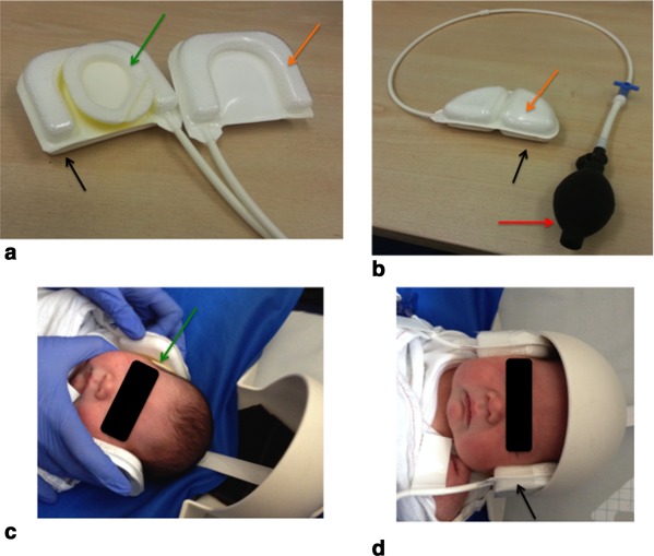
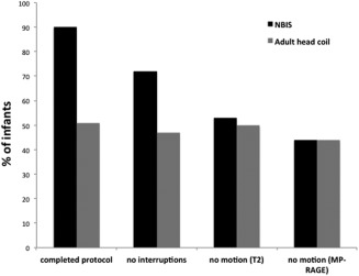
Methods: We designed a neonatal brain imaging system (NBIS) consisting of a dedicated 32-channel receive array coil and a positioning device that allows placement of the infant's head deep into the coil for maximum signal-to-noise ratio (SNR). Disturbance to the infant was minimized by using an MRI-compatible trolley to prepare and transport the infant and by employing a slow ramp-up and continuation of gradient noise during scanning. Scan repeats were minimized by using a restart capability for diffusion MRI and retrospective motion correction. We measured the 1) SNR gain, 2) number of infants with a completed scan protocol, and 3) number of anatomical images with no motion artifact using NBIS compared with using an adult 32-channel head coil.

Results: The NBIS has 2.4 times the SNR of the adult coil and 90% protocol completion rate.

Conclusion: The NBIS allows advanced neonatal brain imaging techniques to be employed in neonatal brain imaging with high protocol completion rates. Magn Reson Med 78:794-804, 2017. © 2016 The Authors Magnetic Resonance in Medicine published by Wiley Periodicals, Inc. on behalf of International Society for Magnetic Resonance in Medicine. This is an open access article under the terms of the Creative Commons Attribution-NonCommercial-NoDerivs License, which permits use and distribution in any medium, provided the original work is properly cited, the use is non-commercial and no modifications or adaptations are made.

Keywords: brain; head coil; neonatal; unsedated.


Figures

Figure 1
Figure 1
MPRAGE midsagittal (a) and coronal (b) views of the neonatal brain. AP measurements were taken from the nasion to the occiput on the midsagittal slice as shown by the horizontal blue line in panel a, and an IS measurement was taken from the top of the skull to the body of the third cervical vertebra as shown by the vertical blue line in the midsagittal slice (a). RL head size was estimated by measuring the biparietal distance (b, blue horizontal line).
Figure 2
Figure 2
Three components of the NBIS, consisting of (a(i)) the neonatal head coil, (a(ii)) the frame, and (a(iii)) the positioning shell. (b) End view of the neonatal head coil showing the AP and RL diameter of the coil (black arrows). (c) Top view of the coil. The black arrow denotes the IS length of the coil; the green arrow indicates a 2‐cm2 gap for respiratory aids; (df) Positioning shell, consisting of a v‐shaped base and a round headpiece with a pocket for the saturation monitor (d, green arrow). The base of the shell has grooves that allows it to be placed securely on the rails of the frame (e, red arrow). The headpiece has three openings for positioning and viewing the final position of the infant's head (e). (f) The distance between the headpiece and the coil is <5 mm to preserve SNR. (h) NBIS system on the scanner table top. (i) Acoustic hood in situ over the NBIS before positioning the infant in the scanner.
Figure 3
Figure 3
Specially designed positioning and immobilization devices. (a, b) The devices consist of bead‐filled cushions (orange arrows) backed by an inflatable air cushion (black arrows). Immobilization devices that sit over the ear muffs (a, green arrow) with an air pump (b, red arrow) allow fine control of head position and comfort. A positioning cushion (b) sits under the base of the neck of the infant. Inflation of this cushion allows fine control of the AP tilt of the infant's head. (c) Positioning of the ear immobilizers over the mini‐muffs (green arrow). (d) Immobilizers in situ with the inflated air pocket (black arrow).
Figure 4
Figure 4
NBIS MRI‐compatible trolley (a) with the shell in situ (b). (a(i)) The green circle and green arrow indicate a close‐up of the breaking system whereby the trolley can only move once the lever is depressed. The rails on which the shell sits is identical to the rails on the NBIS frame that sits on the scanner bed. The frame can be locked (b(i)) and unlocked (b(ii)) to allow the shell to rock backward and forward to settle the infant.
Figure 5
Figure 5
(a) Uniform SNR for a phantom in the adult coil. (b) Enhanced SNR results for the phantom in the neonatal coil. (i, ii) The SNR gain is evident in DTI images performed in term babies with greater contrast‐to‐noise ratio in the cortex for the infant imaged in the neonatal coil (ii) compared with the adult coil (i). (iii) Image of the phantom used to assess the SNR in each coil. A, anterior; I, inferior; L, left; P, posterior, R, right.
Figure 6
Figure 6
Three examples of axial T2 TSE and MPRAGE images uploaded on RView. Example (i) shows no evidence of motion on the acquired T2 axial (i)(a), and there are no disrupted slices in the two orthogonal T2 planes (i)(b) and (i)(c). These images were determined to have no motion. Example (ii) had evidence of minor motion artifact present on the coronal (ii)(b) and sagittal planes (ii)(c). Example (iii) had gross motion artifact on the coronal and sagittal planes (iii)(b) and (iii)(c) and axial MPRAGE (iii)(d). Motion evidenced by disrupted slices in the T2 orthogonal views are indicated by the green arrows. Example of no motion on MPRAGE were as in example (i)(d), moderate motion in row (ii)(d), and gross motion in row (iii)(d).
Figure 7
Figure 7
Performance of the NBIS compared with the adult head coil. The neonatal system had a greater number of infants with a completed protocol and fewer interruptions due to the infant waking than using the adult coil. There was little to no difference in the number of images with evidence of motion artifact on T2 TSE and MPRAGE images.

References

    1. Mathur AM, Neil JJ, McKinstry RC, Inder TE. Transport, monitoring, and successful brain MR imaging in unsedated neonates. Pediatr Radiol 2008;38:260–264. - PubMed
    1. Ureta‐Velasco N, Martinez‐de Aragon A, Moral‐Pumarega MT, Nunez‐Enamorado N, Bergon‐Sendin E, Pallas‐Alonso CR. Magnetic resonance imaging without sedation in neonates [in Spanish]. An Pediatr (Barc) 2015;82:354–359. - PubMed
    1. Keil B, Alagappan V, Mareyam A, et al. Size‐optimized 32‐channel brain arrays for 3T pediatric imaging. Magn Reson Med 2011;66:1777–1787. - PMC - PubMed
    1. Tocchio S, Kline‐Fath B, Kanal E, Schmithorst VJ, Panigrahy A. MRI evaluation and safety in the developing brain. Semin Perinatol 2015;39:73–104. - PMC - PubMed
    1. Golan A, Marco R, Raz H, Shany E. Imaging in the newborn: infant immobilizer obviates the need for anesthesia. Isr Med Assoc J 2011;13:663–665. - PubMed

Publication types

MeSH terms