. 2016 Sep 19;11(9):e0162398.
doi: 10.1371/journal.pone.0162398. eCollection 2016.

Disruption of Prostate Microvasculature by Combining Microbubble-Enhanced Ultrasound and Prothrombin

Affiliations

Disruption of Prostate Microvasculature by Combining Microbubble-Enhanced Ultrasound and Prothrombin

Jinlong Zhang et al. PLoS One. .

Abstract

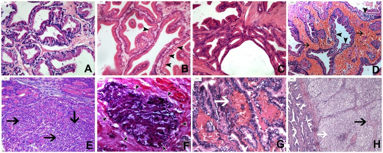
Previous studies have shown a unique method to disrupt tumor vasculature using pulsed, high-pressure amplitude therapeutic ultrasound combined with microbubbles. In this study, we attempted to destroy the prostate vasculature of canine prostates using microbubble-enhanced ultrasound (MEUS) and prothrombin. The prostates of 43 male mongrel canines were surgically exposed. Twenty-two prostates were treated using MEUS (n = 11) or MEUS and prothrombin (PMEUS, n = 11). The other 21 prostates, which were treated using microbubbles (n = 7), ultrasound (n = 7) or prothrombin (n = 7) only, served as the controls. Prothrombin was intravenously infused at 20 IU/kg. MEUS was induced using a therapeutic ultrasound device at a peak negative pressure of 4.47 MPa and a microbubble injection. Contrast-enhanced ultrasound was performed to assess the blood perfusion of the prostates. Then, the prostate tissue was harvested immediately after treatment and at 48 hours later for pathological examination. The contrast-enhanced ultrasound peak value of the prostate decreased significantly from 36.2 ± 5.6 to 27.1 ± 6.3 after treatment in the PMEUS group, but it remained unchanged in the other groups. Histological examination found severe microvascular rupture, hemorrhage and thrombosis in both MEUS- and PMEUS-treated prostates immediately after treatment, while disruption in the PMEUS group was more severe than in the MEUS group. Forty-eight hours after treatment, massive necrosis and infiltration of white blood cells occurred in the PMEUS group. This study demonstrated that PMEUS disrupted the normal microvasculature of canine prostates and induced massive necrosis. PMEUS could potentially become a new noninvasive method used for the treatment of benign prostatic hyperplasia.


Conflict of interest statement

The authors have declared that no competing interests exist.

Figures

Fig 1
Fig 1. Analysis of contrast-enhanced ultrasound in the microbubble-enhanced ultrasound group.
The upper panels in A (before treatment) and B (after treatment) showed the contrast-enhanced ultrasound image with two modes: contrast mode (left) and tissue mode (right). A rectangular region of interest was defined inside the frontal prostate, excluding the acoustically attenuated area. The lower panels showed a time intensity curve (TIC) generated automatically by QLAB software. (A) After a rapid ascension in acoustic intensity, the TIC showed a relative plateau around peak intensity (representing microbubble diffusion to the mesenchyme) and then a slow descent. (B) The ascension and descension of the TIC after treatment were steeper, and the diffusion plateau became imperceptible or even disappeared.
Fig 2
Fig 2. Analysis of the contrast-enhanced ultrasound in microbubble-enhanced ultrasound and prothrombin treated prostates.
Interface of the acoustic quantification software showing the contrast-enhanced ultrasound and the 2-D images. (A) A spherical region of interest was set, and a time-intensity curve was analyzed before treatment. (B) In the microbubble-enhanced ultrasound and prothrombin groups, the curves after treatment show that the value of the peak decreased, compared with before treatment.
Fig 3
Fig 3. Contrast-enhanced ultrasound images of the microbubble-enhanced ultrasound and prothrombin and prothrombin-treated prostates.
(B) The microbubble-enhanced ultrasound and prothrombin-treated prostate, demonstrating a significant blood perfusion decrease in the targeted area. (A, C, D) The baseline in the microbubble-enhanced ultrasound and prothrombin group (A) and all of the corresponding contrast-enhanced ultrasound images (C: before treatment, D: after treatment) of the prothrombin-treated prostates, showing homogenous blood perfusion.
Fig 4
Fig 4. Prostate samples of the different groups obtained after treatment (scale bar = 5 mm).
(A-C) Pictures from the prothrombin, microbubbles, and ultrasound-treated specimens (A: Prothrombin group, B: MB group, C: US group) demonstrated normal prostate tissue without hemorrhage. (D, E) The microbubble-enhanced ultrasound (MEUS)-treated prostate showed severe hemorrhage, especially in the tissue surrounding the urethra (D: severe hemorrhage immediately after treatment, E: hemorrhage with irregularly shaped and blurred boundaries 48 hours later). (F, G) The microbubble-enhanced ultrasound and prothrombin (PMEUS)-treated prostate showed more gross severe hemorrhage than the MEUS group throughout most of the gland, and the color was darker (F: severe hemorrhage immediately after treatment, G: diffused hemorrhage with irregular shape 48 hours later). (H) Coronal plains of the prostates with a dividing line of the urinary track demarcating the two parts after Evans blue dyeing. In the presence of microbubbles and prothrombin, the control portion (left) without ultrasound irradiation was homogeneously dyed blue. The ultrasound irradiated part (right) was dark red with severe hemorrhage, indicating that the Evans blue stain was blocked by PMEUS.
Fig 5
Fig 5. Prostate samples harvested from different groups after treatment (scale bar = 25 μm in A-G and 100 μm in H).
(A) Normal histological structures displayed in the MB group. (B) Vacuolization of epithelial cells [arrowhead] was displayed in the US group immediately after ultrasound irradiation. (C) Almost complete recovery of the affected cells 48 hours later in the US group. (D-F) Microscopic examination results after microbubble-enhanced ultrasound (MEUS) treatment (D: Vacuolization of epithelial cells [arrowhead], red blood cells filling the stroma [arrow], diffuse thrombosis, enlarging and squeezing of the stroma immediately after MEUS; E: Areas of diffuse neutrophil infiltration [arrow] were observed frequently in the stroma and glandular lumen 48 hours after treatment; F: Collapse of glandular structure [arrowhead] and focal necrosis with pyknosis 4 days after treatment). (G, H) Microscopic examination results after microbubble-enhanced ultrasound and prothrombin (PMEUS) treatment (G: More severe hemorrhage and thrombosis than the MEUS group in the interstitial tissues immediately after PMEUS [white arrow]; H: Significant squeezing and focal necrosis of prostatic tissues [black arrow] by the compression of neutrophil infiltration 48 hours after treatment [white arrow].

References

    1. Cabelin MA, Te AE, Kaplan SA. Benign prostatic hyperplasia: Challenges for the new millennium. Curr Opin Urol. 2000; 10: 301–306. - PubMed
    1. Yu X, Elliott SP, Wilt TJ, McBean AM. Practice patterns in benign prostatic hyperplasia surgical therapy: the dramatic increase in minimally invasive technologies. J Urol. 2008; 180: 241–245. 10.1016/j.juro.2008.03.039 - DOI - PubMed
    1. Tunuguntla HS, Evans CP. Minimally invasive therapies for benign prostatic hyperplasia. World J Urol. 2002; 20:197–206. - PubMed
    1. Ohigashi T, Nakamura K, Nakashima J, Baba S, Murai M. Long-term results of three different minimally invasive therapies for lower urinary tract symptoms due to benign prostatic hyperplasia: comparison at a single institute. Int J Urol. 2007; 14: 326–330. - PubMed
    1. McNeal JE. Origin and evolution of benign prostatic enlargement. Invest Urol. 1978; 15: 340–345. - PubMed