. 2016 Dec;15(12):3581-3593.
doi: 10.1074/mcp.M116.060806. Epub 2016 Sep 19.

Identification of 2-oxohistidine Interacting Proteins Using E. coli Proteome Chips

Affiliations

Identification of 2-oxohistidine Interacting Proteins Using E. coli Proteome Chips

Jun-Mu Lin et al. Mol Cell Proteomics. 2016 Dec.

Abstract

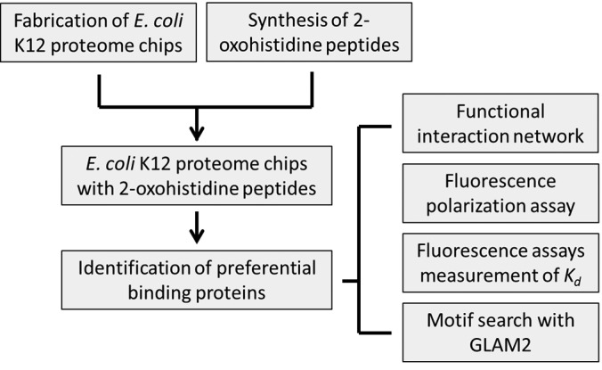
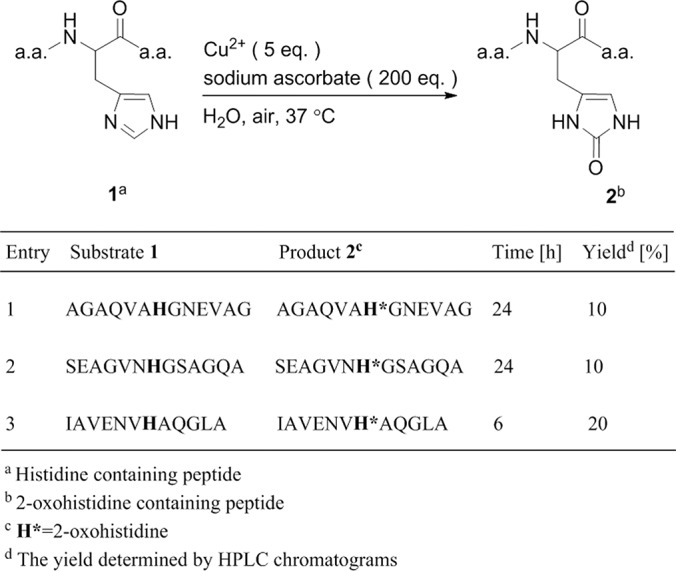
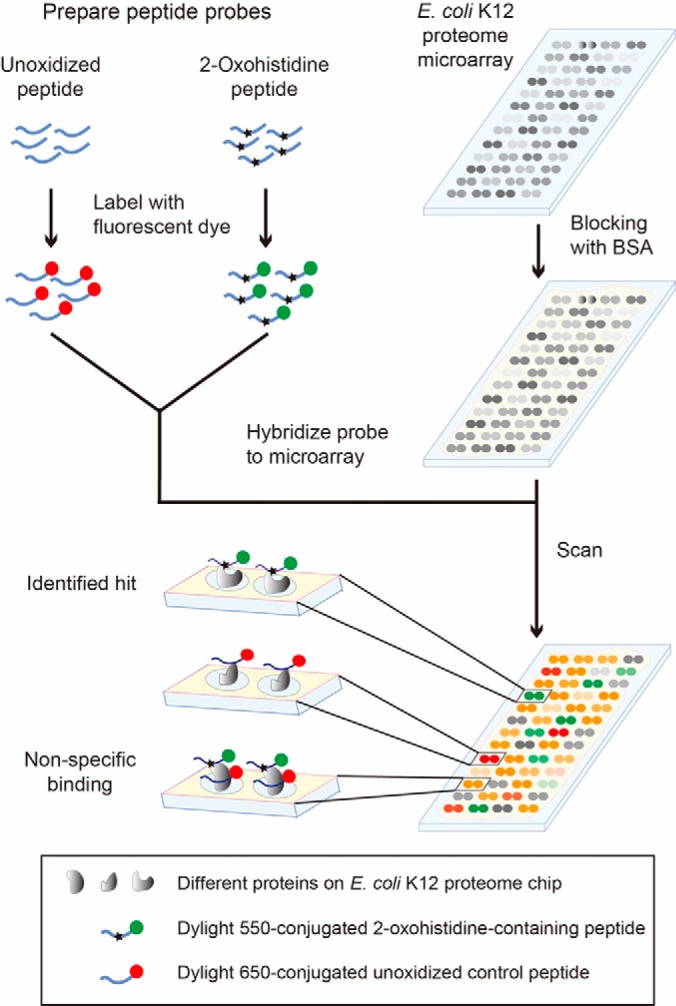
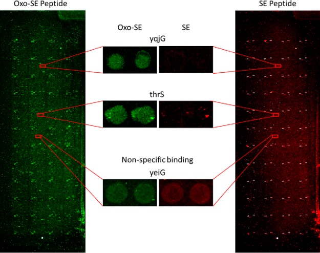
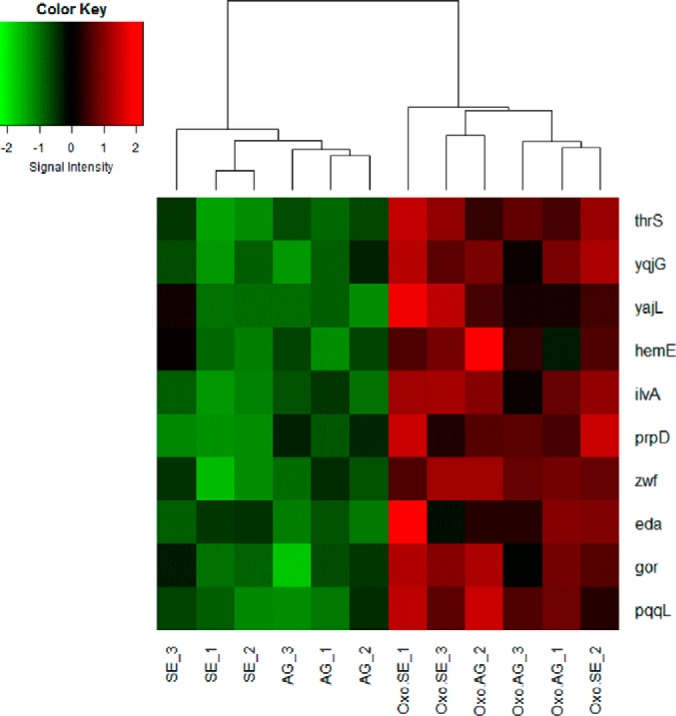
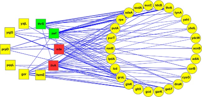
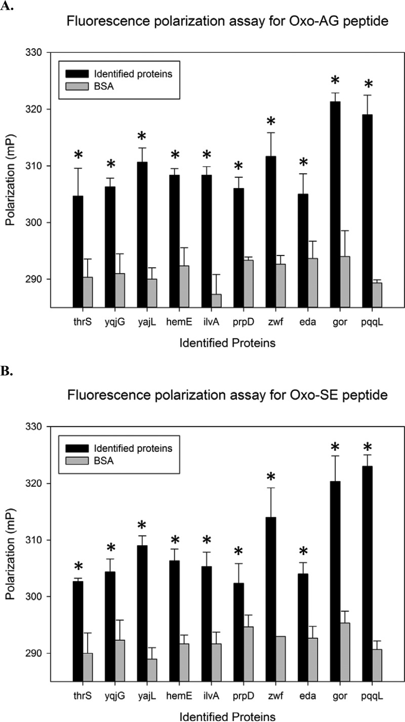
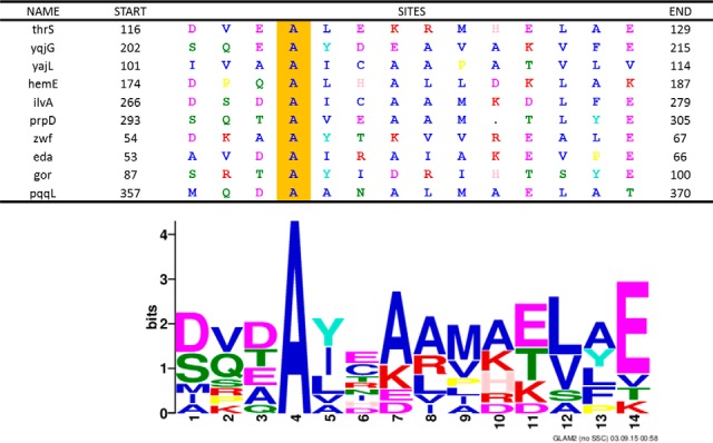
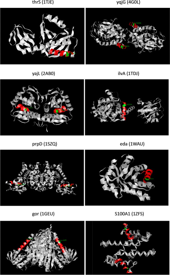
Cellular proteins are constantly damaged by reactive oxygen species generated by cellular respiration. Because of its metal-chelating property, the histidine residue is easily oxidized in the presence of Cu/Fe ions and H2O2 via metal-catalyzed oxidation, usually converted to 2-oxohistidine. We hypothesized that cells may have evolved antioxidant defenses against the generation of 2-oxohistidine residues on proteins, and therefore there would be cellular proteins which specifically interact with this oxidized side chain. Using two chemically synthesized peptide probes containing 2-oxohistidine, high-throughput interactome screening was conducted using the E. coli K12 proteome microarray containing >4200 proteins. Ten interacting proteins were identified, and successfully validated using a third peptide probe, fluorescence polarization assays, as well as binding constant measurements. We discovered that 9 out of 10 identified proteins seemed to be involved in redox-related cellular functions. We also built the functional interaction network to reveal their interacting proteins. The network showed that our interacting proteins were enriched in oxido-reduction processes, ion binding, and carbon metabolism. A consensus motif was identified among these 10 bacterial interacting proteins based on bioinformatic analysis, which also appeared to be present on human S100A1 protein. Besides, we found that the consensus binding motif among our identified proteins, including bacteria and human, were located within α-helices and faced the outside of proteins. The combination of chemically engineered peptide probes with proteome microarrays proves to be an efficient discovery platform for protein interactomes of unusual post-translational modifications, and sensitive enough to detect even the insertion of a single oxygen atom in this case.


Figures

Fig. 1.
Fig. 1.
Overall strategy for the identification of 2-oxohistidine interacting proteins using E. coli K12 proteome chip. We expressed and purified ∼4,300 E. coli proteins in high-throughput manner to fabricate the E. coli K12 proteome chip. We used an improved condition to obtain 2-oxohistidine peptides in high yield and purity. 2-Oxohistidine peptides were then probed to E. coli K12 proteome chip and preferential binding proteins were identified. We also built their functional interaction network to investigate their biological roles. Fluorescence polarization assays were used to validate the identified binding proteins. We conducted dose-response fluorescence assays to measure the Kd of these proteins. Furthermore, we used GLAM2 to search consensus motif among these identified proteins and also applied this motif to the entire E. coli K12 proteome and human proteome by GLAM2SCAN.
Fig. 2.
Fig. 2.
Summary scheme for the synthesis of 2-oxohistidine-containing peptides. Using metal-catalyzed oxidation, the histidine side chain on three different peptides (AG, SE, IA) was converted to 2-oxohistidine.
Fig. 3.
Fig. 3.
Schematic of E. coli K12 proteome chip assays with 2-oxohistidine peptide probes. To detect 2-oxohistidine interacting proteins, E. coli K12 proteome chips were probed with 2-oxohistidine-containing peptides and unoxidized control peptides labeled with different fluorophores. Each protein was printed in duplicate spots on the chip.
Fig. 4.
Fig. 4.
Representative images of the E. coli K12 proteome chips probed with 2-oxohistidine containing peptide (Oxo-SE peptide) and unoxidized peptide (SE peptide). The representative positive hits (yqjG and thrS) and nonspecific binding protein (yeiG) on the chip are enlarged from sample images of Oxo-SE peptide and SE peptide, respectively. The contrast and brightness of images have been adjusted equally using the same parameters.
Fig. 5.
Fig. 5.
The heat map of 10 identified proteins. The heat map shows the classification of 10 identified proteins in Oxo-AG, Oxo-SE, AG, and SE chip assay probing results. Each peptide probe were analyzed in triplicate. The R programming language and gplots package were used to display the heat map.
Fig. 6.
Fig. 6.
The functional interaction network of the 10 identified 2-oxohistidine binding proteins and 26 secondary interacting proteins. The interaction pairs for 10 identified proteins were downloaded from EcID database, and functional interaction network was visualized by Cytoscape. We found 26 secondary interacting proteins that interacted with at least three out of the 10 proteins which bind 2-oxohistidine. Four out of ten identified proteins, (eda, ilvA, zwf, and thrS) have many interactions and were considered to be hubs. Square shapes represent the 10 identified proteins, and round shapes represent the 26 secondary interacting proteins. The node colors reflect the number of interactions: the red for >10 interactions, the green for 5–10 interactions, and the yellow for <5 interactions. The thickness of connecting lines reflects the strength of protein-protein interactions, determined by the number of reported interactions from database searches.
Fig. 7.
Fig. 7.
Validation of the interactions between 2-oxohistidine peptides and identified proteins using fluorescence polarization assays. Tested proteins were compared with identical concentrations of BSA, which served as the negative control. A, The fluorescence polarization assays for Oxo-AG peptide and identified proteins. B, The fluorescence polarization assays for Oxo-SE peptide and identified proteins. The error bars represent S.E. The asterisks indicate p < 0.05 by unpaired t test against the BSA control.
Fig. 8.
Fig. 8.
Consensus motif among the 10 validated proteins. The logo shows the consensus motif [DS][VQ][DTE]A[YIL]X[AK][ARL][MV][ETK][LV][AYLF]E identified by GLAM2. The table shows the protein sequences of 10 validated 2-oxohistidine binding proteins aligned with the consensus motif.
Fig. 9.
Fig. 9.
Protein 3D structures of E. coli identified proteins and human S100A1. Seven of the E. coli identified proteins (thrS, yqjG, yajL, ilvA, prpD, eda, gor) and human S100A1 have published protein 3D structures, which are visualized by RasMol software. The red region represents the consensus motif found by GLAM2, and the green region indicates the conserved alanine at the fourth position in the consensus motif. YqjG, yajL, prpD, gor and S100A1 were deposited in RCSB PDB as homodimer structures, whereas the others were monomers.

References

    1. Wold F. (1981) In vivo chemical modification of proteins (post-translational modification). Annu. Rev. Biochem. 50, 783–814 - PubMed
    1. Walsh C. T., Garneau-Tsodikova S., and Gatto G. J. Jr. (2005) Protein posttranslational modifications: the chemistry of proteome diversifications. Angew Chem. Int. Ed. 44, 7342–7372 - PubMed
    1. Harding J. J. (1985) Nonenzymatic covalent posttranslational modification of proteins in vivo. Adv. Protein Chem. 37, 247–334 - PubMed
    1. Davies M. J. (2005) The oxidative environment and protein damage. Biochim. Biophys. Acta 1703, 93–109 - PubMed
    1. Morrison D. K. (2009) The 14-3-3 proteins: integrators of diverse signaling cues that impact cell fate and cancer development. Trends Cell Biol. 19, 16–23 - PMC - PubMed

LinkOut - more resources