

Ocular indicators of Alzheimer's: exploring disease in the retina
- PMID: 27645291
- PMCID: PMC5106496
- DOI: 10.1007/s00401-016-1613-6
Ocular indicators of Alzheimer's: exploring disease in the retina
Abstract
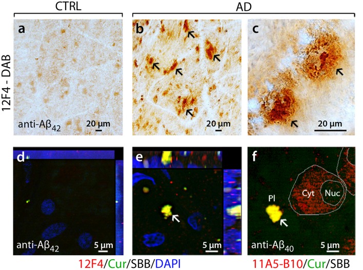
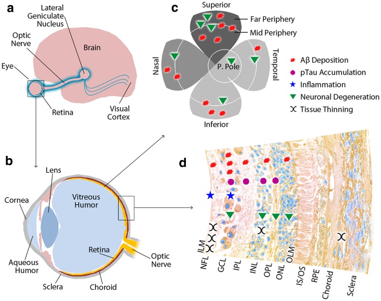
Although historically perceived as a disorder confined to the brain, our understanding of Alzheimer's disease (AD) has expanded to include extra-cerebral manifestation, with mounting evidence of abnormalities in the eye. Among ocular tissues, the retina, a developmental outgrowth of the brain, is marked by an array of pathologies in patients suffering from AD, including nerve fiber layer thinning, degeneration of retinal ganglion cells, and changes to vascular parameters. While the hallmark pathological signs of AD, amyloid β-protein (Aβ) plaques and neurofibrillary tangles (NFT) comprising hyperphosphorylated tau (pTau) protein, have long been described in the brain, identification of these characteristic biomarkers in the retina has only recently been reported. In particular, Aβ deposits were discovered in post-mortem retinas of advanced and early stage cases of AD, in stark contrast to non-AD controls. Subsequent studies have reported elevated Aβ42/40 peptides, morphologically diverse Aβ plaques, and pTau in the retina. In line with the above findings, animal model studies have reported retinal Aβ deposits and tauopathy, often correlated with local inflammation, retinal ganglion cell degeneration, and functional deficits. This review highlights the converging evidence that AD manifests in the eye, especially in the retina, which can be imaged directly and non-invasively. Visual dysfunction in AD patients, traditionally attributed to well-documented cerebral pathology, can now be reexamined as a direct outcome of retinal abnormalities. As we continue to study the disease in the brain, the emerging field of ocular AD warrants further investigation of how the retina may faithfully reflect the neurological disease. Indeed, detection of retinal AD pathology, particularly the early presenting amyloid biomarkers, using advanced high-resolution imaging techniques may allow large-scale screening and monitoring of at-risk populations.
Keywords: Alzheimer’s disease; Amyloid-beta; Neurodegenerative disease; Ocular abnormalities; Retinal biomarkers; Tauopathy.
Conflict of interest statement
YK, MKH, KLB, founding members of NeuroVision Imaging (NVI).
Figures
References
-
- Algvere PV, Kvanta A, Seregard S. Drusen maculopathy: a risk factor for visual deterioration. Acta Ophthalmol. 2016;94:427–433. - PubMed
Publication types
MeSH terms
Substances
Grants and funding
LinkOut - more resources
Full Text Sources
Other Literature Sources
Medical
Research Materials
