Review
. 2016 Sep 20:14:271.
doi: 10.1186/s12967-016-1028-0.

Recent advances in bioprinting techniques: approaches, applications and future prospects

Affiliations
Review

Recent advances in bioprinting techniques: approaches, applications and future prospects

Jipeng Li et al. J Transl Med. .

Abstract

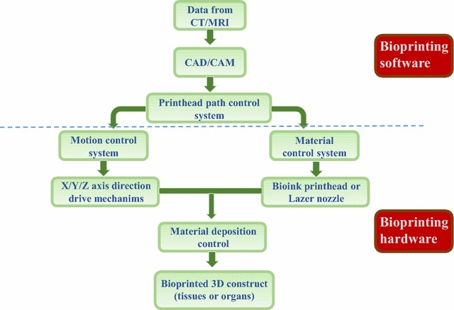
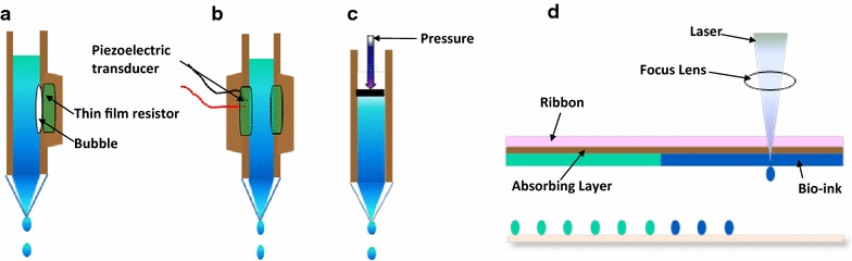
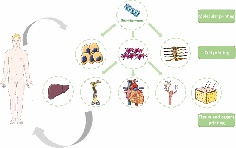
Bioprinting technology shows potential in tissue engineering for the fabrication of scaffolds, cells, tissues and organs reproducibly and with high accuracy. Bioprinting technologies are mainly divided into three categories, inkjet-based bioprinting, pressure-assisted bioprinting and laser-assisted bioprinting, based on their underlying printing principles. These various printing technologies have their advantages and limitations. Bioprinting utilizes biomaterials, cells or cell factors as a "bioink" to fabricate prospective tissue structures. Biomaterial parameters such as biocompatibility, cell viability and the cellular microenvironment strongly influence the printed product. Various printing technologies have been investigated, and great progress has been made in printing various types of tissue, including vasculature, heart, bone, cartilage, skin and liver. This review introduces basic principles and key aspects of some frequently used printing technologies. We focus on recent advances in three-dimensional printing applications, current challenges and future directions.

Keywords: 3D bioprinting; Artificial organs; Tissue engineering.


Figures

Fig. 1
Fig. 1
General 3D bioprinting technical route
Fig. 2
Fig. 2
Common types of bioprinting methods. a Thermal inkjet-based bioprinting technology utilizes an electric current pulse that impulses the thin film resistor, then generates bubbles that create a pressure pulse that propels the ink droplet onto the substrates. b A piezoelectric transducer creates a pulse that creates transient pressure, resulting in droplet ejection. c Pressure-assisted bioprinting uses solutions, pastes, or dispersions as biomaterials, which are extruded by pressure in the form of a continuous filament through a microscale nozzle orifice or a microneedle. d Laser-associated bioprinting consists of three parts: a pulsed laser source, a ribbon and a receiving substrate. The lasers irradiate the ribbon, causing the liquid biological materials to evaporate and reach the receiving substrate in droplet form
Fig. 3
Fig. 3
The applications of bioprinting range from the molecular level to organ level

References

    1. Huang JMY, Millis JM. Government policy and organ transplantation in China. Lancet. 2008;372:1937–1938. doi: 10.1016/S0140-6736(08)61359-8. - DOI - PubMed
    1. Zhang Q, Johnson JA, Dunne LW, Chen Y, Iyyanki T, Wu Y, Chang EI, Branch-Brooks CD, Robb GL, Butler CE. Decellularized skin/adipose tissue flap matrix for engineering vascularized composite soft tissue flaps. Acta Biomater. 2016;35:166–184. doi: 10.1016/j.actbio.2016.02.017. - DOI - PMC - PubMed
    1. Futrega K, Palmer JS, Kinney M, Lott WB, Ungrin MD, Zandstra PW, Doran MR. The microwell-mesh: a novel device and protocol for the high throughput manufacturing of cartilage microtissues. Biomaterials. 2015;62:1–12. doi: 10.1016/j.biomaterials.2015.05.013. - DOI - PubMed
    1. Shamaz BH, Anitha A, Vijayamohan M, Kuttappan S, Nair S, Nair MB. Relevance of fiber integrated gelatin-nanohydroxyapatite composite scaffold for bone tissue regeneration. Nanotechnology. 2015;26:405101. doi: 10.1088/0957-4484/26/40/405101. - DOI - PubMed
    1. Lee CH, Lee FY, Tarafder S, Kao K, Jun Y, Yang G, Mao JJ. Harnessing endogenous stem/progenitor cells for tendon regeneration. J Clin Invest. 2015;125:2690–2701. doi: 10.1172/JCI81589. - DOI - PMC - PubMed

Publication types

Substances