. 2016 Dec;139(Pt 12):3151-3162.
doi: 10.1093/brain/aww229. Epub 2016 Sep 19.

Microbleed and microinfarct detection in amyloid angiopathy: a high-resolution MRI-histopathology study

Affiliations

Microbleed and microinfarct detection in amyloid angiopathy: a high-resolution MRI-histopathology study

Susanne J van Veluw et al. Brain. 2016 Dec.

Abstract

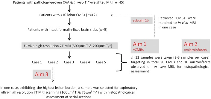
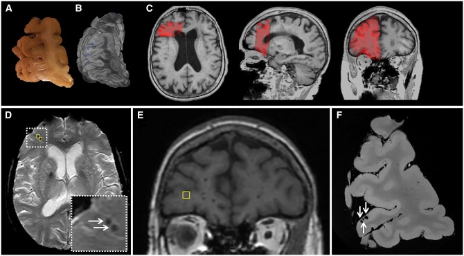
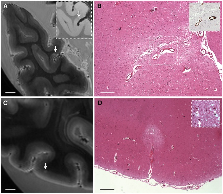
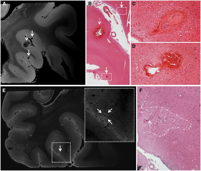
Cerebral amyloid angiopathy is a common neuropathological finding in the ageing human brain, associated with cognitive impairment. Neuroimaging markers of severe cerebral amyloid angiopathy are cortical microbleeds and microinfarcts. These parenchymal brain lesions are considered key contributors to cognitive impairment. Therefore, they are important targets for therapeutic strategies and may serve as surrogate neuroimaging markers in clinical trials. We aimed to gain more insight into the pathological basis of magnetic resonance imaging-defined microbleeds and microinfarcts in cerebral amyloid angiopathy, and to explore the pathological burden that remains undetected, by using high and ultra-high resolution ex vivo magnetic resonance imaging, as well as detailed histological sampling. Brain samples from five cases (mean age 85 ± 6 years) with pathology-proven cerebral amyloid angiopathy and multiple microbleeds on in vivo clinical magnetic resonance imaging were subjected to high-resolution ex vivo 7 T magnetic resonance imaging. On the obtained high-resolution (200 μm isotropic voxels) ex vivo magnetic resonance images, 171 microbleeds were detected compared to 66 microbleeds on the corresponding in vivo magnetic resonance images. Of 13 sampled microbleeds that were matched on histology, five proved to be acute and eight old microhaemorrhages. The iron-positive old microhaemorrhages appeared approximately four times larger on magnetic resonance imaging compared to their size on histology. In addition, 48 microinfarcts were observed on ex vivo magnetic resonance imaging in three out of five cases (two cases exhibited no microinfarcts). None of them were visible on in vivo 1.5 T magnetic resonance imaging after a retrospective analysis. Of nine sampled microinfarcts that were matched on histology, five were confirmed as acute and four as old microinfarcts. Finally, we explored the proportion of microhaemorrhage and microinfarct burden that is beyond the detection limits of ex vivo magnetic resonance imaging, by scanning a smaller sample at ultra-high resolution, followed by serial sectioning. At ultra-high resolution (75 μm isotropic voxels) magnetic resonance imaging we observed an additional 48 microbleeds (compared to high resolution), which proved to correspond to vasculopathic changes (i.e. morphological changes to the small vessels) instead of frank haemorrhages on histology. After assessing the serial sections of this particular sample, no additional haemorrhages were observed that were missed on magnetic resonance imaging. In contrast, nine microinfarcts were found in these sections, of which six were only retrospectively visible at ultra-high resolution. In conclusion, these findings suggest that microbleeds on in vivo magnetic resonance imaging are specific for microhaemorrhages in cerebral amyloid angiopathy, and that increasing the resolution of magnetic resonance images results in the detection of more 'non-haemorrhagic' pathology. In contrast, the vast majority of microinfarcts currently remain under the detection limits of clinical in vivo magnetic resonance imaging.

Keywords: histology; microbleeds; microinfarcts; post-mortem MRI; small vessel disease.


Figures

Figure 1
Figure 1
Study design and flowchart. Twelve of 45 patients from the whole cohort had >10 lobar CMBs on their last MRI before death, and were selected for the ex vivo MRI study. Upon retrieval of the stored brain slabs from the local neuropathology database, brain tissue proved to be available for 5 of 12 cases. Hence, intact formalin-fixed brain slabs from five cases were selected for ex vivo MRI scanning. We first assessed CMBs on the obtained ex vivo MRI, followed by detailed histopathological examination of a representative subset of these lesions (Aim 1). In one case with adequate in vivo MRI scan quality, we were able to match CMBs observed on in vivo MRI to the corresponding ex vivo MRI and histopathology sections (sub-Aim 1 b). Second, we assessed microinfarcts on the same ex vivo MRI images, followed by detailed histopathological examination of a representative subset of these lesions (Aim 2). Finally, we rescanned one smaller sample cut from a slab containing the highest number of magnetic resonance-observed CMBs, with a dedicated ultra-high resolution ex vivo MRI protocol, followed by serial sectioning of the whole tissue. Hence we explored the microvascular abnormalities that are visible at this ultra-high resolution, but remained undetected at high resolution ex vivo MRI and in vivo MRI, and studied their underlying histopathology (Aim 3).
Figure 2
Figure 2
Histopathology of representative examples of magnetic resonance-observed microbleeds. (A–C) A cortical microbleed observed on ex vivo T2*-weighted MRI in Case 5 (A; arrow) was sampled for histopathological analysis and matched with a focal accumulation of haemosiderin-containing macrophages on haematoxylin and eosin, representing an old microhaemorrhage (B). The adjacent section was positive for iron (C). Scale bar in A = 4 mm; B and C = 500 µm. (D–F) A cortical microbleed observed on ex vivo T2*-weighted MRI in Case 2 (D; arrow) was sampled for histopathological analysis and matched with a focal accumulation of erythrocytes close to a ruptured vessel, representing an acute microhaemorrhage (E). The adjacent section was partly positive for iron, suggesting that the same vessel had ruptured before (F). Scale bar in D = 4 mm; E and F = 250 µm.
Figure 3
Figure 3
Matched microbleeds between in vivo and ex vivo MRI, after registration. From one case the tissue volume that was subjected to ex vivo MRI (A and B) could be matched with the corresponding in vivo MRI, acquired 1 year and 14 days ante-mortem (C). First, the ex vivo T2*-weighted MRI (B) was matched to the in vivo 3D T1-weighted 1.5 T MRI, by means of manual and linear registration (C). B represents a volume rendering of the four continuous slabs taken from the right frontal area stacked on top of each other. The blue dots represent microbleeds observed on ex vivo MRI. Second, observed microbleeds on the in vivo T2*-weighted 1.5 T MRI (D) were registered to the coronal in vivo T1-weighted MRI image (E; marker corresponds to left marker in D) and compared to the volume-matched ex vivo T2*-weighted MRI image (F). Hence, all seven microbleeds that were found on in vivo MRI in this case could be matched to microbleeds on the corresponding ex vivo MRI. Three microbleeds, which were both identified on in vivo (D, two are captured in this transversal view) and ex vivo MRI (F, all three are captured in this coronal view), were subsequently sampled for histopathological examination and proved to be old microhaemorrhages on microscopy.
Figure 4
Figure 4
Histopathology of representative examples of magnetic resonance-observed microinfarcts. (A and B) A cortical microinfarct observed on ex vivo T2-weighted MRI in Case 3 (A; arrow; inset is T2*-weighted MRI) was sampled for histopathological analysis and matched with a region of pallor, tissue loss, and gliosis on haematoxylin and eosin, representing a chronic microinfarct (B). The inset in B shows amyloid-β positive cortical vessels associated with the microinfarct, observed on an adjacent section stained for amyloid-β. Scale bars in A = 4 mm; B = 250 µm. (C and D) A cortical microinfarct observed on ex vivo T2-weighted MRI in Case 2 (C; arrow) was sampled for histopathological analysis and matched with a region of tissue pallor containing ischaemic neurons (arrows; inset), representing an acute microinfarct (D). Scale bars in C = 4 mm; D = 500 µm.
Figure 5
Figure 5
Exploratory ultra-high resolution ex vivo MRI. Ultra-high resolution ex vivo magnetic resonance images of a sampled brain area from Case 2 reveal striking detail of CAA-related pathology. Top row: Here we show three representative microbleeds that were identified on the ultra-high resolution T2*-weighted ex vivo magnetic resonance image (voxel size 75 µm3), of which the larger one (broken arrow) was also visible at the corresponding high resolution T2*-weighted ex vivo magnetic resonance image (voxel size 200 µm3) (A). This microbleed corresponded to a recent microhaemorrhage on haematoxylin and eosin, characterized by a focal accumulation of intact erythrocytes (broken arrow; B). The hypointense lesions (arrows) that were not rated as microbleeds at high-resolution T2*-weighted ex vivo MRI, proved to be vasculopathies on haematoxylin and eosin, without parenchymal tissue injury (arrows; B, enlarged in C and D). The vasculopathy in C resembles an occluded vessel containing fibrin deposits. The vasculopathy in D resembles a microaneurysm. Bottom row: Here we show a microinfarct that was identified on microscopic examination of the serial histological sections taken from this sample (F), and retrospectively could be identified as a hyperintense lesion on the corresponding ultra-high resolution T2-weighted ex vivo magnetic resonance image (voxel size 100 µm3) (E), whereas it escaped detection at high-resolution T2-weighted ex vivo MRI (voxel size 300 µm3). Note the vessel at the centre of this microinfarct (broken arrow in F), which can be distinguished on the scan (hypointense structure within the hyperintense lesion in E).

References

    1. Arvanitakis Z, Leurgans SE, Wang Z, Wilson RS, Bennett DA, Schneider JA. Cerebral amyloid angiopathy pathology and cognitive domains in older persons .Ann Neurol 2011a; 69: 320–7. - PMC - PubMed
    1. Arvanitakis Z, Leurgans SE, Barnes LL, Bennett DA, Schneider JA. Microinfarct pathology, dementia, and cognitive systems. Stroke 2011b; 42: 722–7. - PMC - PubMed
    1. Boyle PA, Yu L, Nag S, Leurgans S, Wilson RS, Bennett DA, et al. Cerebral amyloid angiopathy and cognitive outcomes in community-based older persons. Ann Neurol 2015; 85: 1930–6. - PMC - PubMed
    1. Brundel M, Heringa SM, de Bresser J, Koek HL, Zwanenburg JJ, Kappelle JL, et al. High prevalence of cerebral microbleeds at 7Tesla MRI in patients with early Alzheimer’s disease. J Alzheimers Dis 2012; 31: 259–63. - PubMed
    1. Charidimou AC, Martinez-Ramirez S, Reijmer YD, Oliveira-Filho J, Lauer A, Roongpiboonsopit D, et al. Total MRI small vessel disease burden in cerebral amyloid angiopathy: a concept validation imaging-pathological study. JAMA Neurol 2016; 73: 994–1001. - PMC - PubMed

Publication types