. 2017 Dec;22(12):1714-1724.
doi: 10.1038/mp.2016.155. Epub 2016 Sep 20.

PDE11A negatively regulates lithium responsivity

Affiliations

PDE11A negatively regulates lithium responsivity

G Pathak et al. Mol Psychiatry. 2017 Dec.

Abstract

Lithium responsivity in patients with bipolar disorder has been genetically associated with Phosphodiesterase 11A (PDE11A), and lithium decreases PDE11A mRNA in induced pluripotent stem cell-derived hippocampal neurons originating from lithium-responsive patients. PDE11 is an enzyme uniquely enriched in the hippocampus that breaks down cyclic AMP and cyclic GMP. Here we determined whether decreasing PDE11A expression is sufficient to increase lithium responsivity in mice. In dorsal hippocampus and ventral hippocampus (VHIPP), lithium-responsive C57BL/6J and 129S6/SvEvTac mice show decreased PDE11A4 protein expression relative to lithium-unresponsive BALB/cJ mice. In VHIPP, C57BL/6J mice also show differences in PDE11A4 compartmentalization relative to BALB/cJ mice. In contrast, neither PDE2A nor PDE10A expression differ among the strains. The compartment-specific differences in PDE11A4 protein expression are explained by a coding single-nucleotide polymorphism (SNP) at amino acid 499, which falls within the GAF-B homodimerization domain. Relative to the BALB/cJ 499T, the C57BL/6J 499A decreases PDE11A4 homodimerization, which removes PDE11A4 from the membrane. Consistent with the observation that lower PDE11A4 expression correlates with better lithium responsiveness, we found that Pde11a knockout mice (KO) given 0.4% lithium chow for 3+ weeks exhibit greater lithium responsivity relative to wild-type (WT) littermates in tail suspension, an antidepressant-predictive assay, and amphetamine hyperlocomotion, an anti-manic predictive assay. Reduced PDE11A4 expression may represent a lithium-sensitive pathophysiology, because both C57BL/6J and Pde11a KO mice show increased expression of the pro-inflammatory cytokine interleukin-6 (IL-6) relative to BALB/cJ and PDE11A WT mice, respectively. Our finding that PDE11A4 negatively regulates lithium responsivity in mice suggests that the PDE11A SNPs identified in patients may be functionally relevant.


Figures
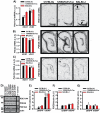
Figure 1
Figure 1. C57BL/6J mice express significantly lower levels of PDE11A4 mRNA than do BALB/cJ mice
To determine if lithium responsiveness may correlate with PDE11A expression across mouse strains, we measured PDE11A4 expression in hippocampi taken from mice that respond well (C57BL/6J), moderately well (129S6/SvEvTac), or poorly to chronic lithium (BALB/cJ). A) PDE11A4 mRNA expression in the C57BL/6J (n=7) and 129S6/SvEvTac mice (n=7) is significantly lower than that in the BALB/cJ mice (n=6); however, this difference in mRNA expression is restricted to dorsal CA1 (DCA1). Data is shown for the oligonucleotide probe that selectively recognizes the PDE11A4 isoform, but is representative of results obtained with 3 different oligonucleotide probes (see Figure S1 for data collected in a second cohort). Surprisingly, BALB/cJ mice fail to show the typical enrichment of PDE11A4 mRNA expression in ventral CA1 (VCA1) vs. DCA1, as has been previously reported in mice and rats and can be seen here in the C57BL/6J mice and 129S6/SvEvTac mice. B) The expression of PDE2A and C) PDE10A mRNA are not significantly different between strains, suggesting specificity of the PDE11A4 effect. D) Western blots measuring PDE11A4 (−control = PDE11A KO VHIPP), PDE2A (− control = cerebellum), PDE10A (+ control = striatum), and actin. E) C57BL/6J and 129S6/SvEvTac mice express significantly less PDE11A4 protein relative to BALB/cJ mice in both DHIPP and VHIPP (n=12/strain). Further, all 3 strains demonstrate far more expression of PDE11A4 protein in VHIPP vs. DHIPP, despite the lack of difference at the mRNA level in the BALB/cJ mice. These effects were replicated in a second cohort of mice (Figure S2). F) Neither PDE2A nor G) PDE10A protein expression differ between the strains, again suggesting specificity of the PDE11A4 effect. Post hoc, * vs BALB/cJ, P<0.001; @vs DCA1 or DHIPP , P<0.017-0.001. R.O.D.—relative optical density. A.U.—arbitrary units. Data pass normality and equal variance and are graphed as means ±SEM. Brightness and contrast adjusted for graphical clarity of autoradiograph and Western blot images.
Figure 2
Figure 2. C57BL/6J mice compartmentalize PDE11A4 protein in VHIPP differently than do BALB/cJ mice
To determine if PDE11A4 protein expression differences between C57BL/6J and BALB/cJ mice might be specific to select subcellular compartments, DHIPP and VHIPP samples were biochemically fractionated and then A) probed by Western Blot. B) C57BL/6J mice showed lower PDE11A4 protein expression relative to BALB/cJ mice in both the cytosolic and membrane compartments of the DHIPP, C) but only in the membrane compartment of the VHIPP. This effect was replicated in a second cohort of mice (Figure S3). DHIPP, n=6/strain for membrane and 8/strain for cytosol; VHIPP, n=8/strain per compartment. Post hoc, *vs BALB/cJ, P<0.01-0.001, #vs cytosol, P<0.001. Data sets passed normality and equal variance and are graphed as means ±SEM. Brightness and contrast adjusted for graphical clarity of blot images.
Figure 3
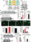
Figure 3. A nonsynonymous coding SNP in the PDE11A4 GAF-B domain promotes PDE11A4 homodimerization and alters PDE11A4 compartmentalization
The PDE11A rs27963339 polymorphism affecting amino acid 499 results in an alanine (499A) in C57BL/6J mice (referred to as wild-type (WT) as this is the sequence published in NCBI) and a threonine (499T) in BALB/cJ mice. A) COS-1 cells were transiently transfected with plasmids encoding an EmGFP-PDE11A4 fusion protein containing a WT 499A, 499T, or 499D (i.e., a phosphomimic aspartate). B) Alternatively, WT PDE11A4 was co-transfected with either mCherry alone or an isolated GAF-B domain fused to mCherry. Hypothetical structure of PDE11A4 based on [81]. C) Co-immunoprecipitation shows that the isolated GAF-B domain binds full-length PDE11A4, enabling the isolated GAF-B domain to act as a dominant negative that prevents homodimerization. D,F) Native PAGE shows that the 499T and 499D mutations increase expression of the presumed homodimer at 252 kDa (n=4/group); whereas, E,G) expression of the isolated GAF-B domain decreases expression of the presumed homodimer (n=6/group). Note: The difference in intensity between the WT vs WT-mCherry groups is not related to the co-expression of mCherry but rather reflects the fact that the experiments were conducted separately and different film exposures were necessary for each experiment in order to keep each group of samples within the linear range of the film (e.g., if WT and WT-mCherry exposures were kept constant between experiments, then the 499 groups would be overexposed). H, I) Consistent with these opposing effects on homodimerization, 499T and 499D promote aggregation of PDE11A4 in COS-1 cells (n=8/group); whereas, J) the isolated GAF-B domain decreases aggregation (n=6/group). K) Importantly, the ability of 499T and 499D to promote PDE11A4 aggregation is blocked when homodimerization is prevented by the isolated GAF-B domain (n=8/group), which suggests the ability of 499T and 499D to increase homodimerization is directly related to their ability to promote the trafficking of PDE11A4 into aggregates. L, N) Biochemical fractionations show that 499T and 499D shift PDE11A4 from the cytosol to the membrane relative to WT PDE11A4 (n=5/group), mimicking PDE11A4 compartmentalization in the VHIPP of BALB/cJ vs. C57BL/6J mice. M,O). In contrast, disrupting homodimerization with the isolated GAF-B domain shifts PDE11A4 from the membrane to the cytosol (mCherry, n=13; GAF-B, n=9). The fact that 499T and 499D behave similarly suggests the 499T construct is naturally phosphorylated in COS-1 cells. Post hoc, *vs. WT, P<0.05-0.001; #vs. 499T, P=0.037-0.011; @vs. mCherry, P<0.001. A.U.—arbitrary units. Note that all n’s reflect biological replicates. Data passed normality and equal variance and are graphed as means ±SEM. Brightness and contrast adjusted for graphical clarity of images.
Figure 4
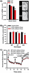
Figure 4. Genetic deletion of PDE11A in mice triggers alterations in the cAMP signaling pathway and increases sensitivity to the antidepressant and anti-manic effects of lithium
A) Autoradiographic in situ hybridization shows that PDE11A KO mice exhibit a significant reduction in Arhgap32 mRNA specifically within ventral CA1 (VCA1) relative to wild-type (WT) littermates (n=9/genotype), indicative of localized increases in pCREB signaling [59, 60]. B) PDE11A KO mice chronically fed 0.4% lithium chow (n=15), but not 0.2% lithium chow (n=21) demonstrated a significant antidepressant-like reduction in tail suspension test (TST) immobility relative to PDE11A KO mice fed a control diet (n=19). PDE11A WT and HT mice showed no significant change in immobility when fed either 0.4% (WT, n=16; HT, n=16) or 0.2% lithium chow (WT, n=20; HT, n=14) relative to the control diet (WT, n=18; HT, n=16). C) At specific time points, PDE11A KO and PDE11A HT mice chronically fed the 0.4% lithium chow demonstrated a significant antimanic-like attenuation of the amphetamine-stimulated rise in horizontal beam breaks relative to WT littermates (WT and HT, n=26; KO, n=25). Post hoc, #vs. control chow, P=0.011; *vs. KO, P=0.015-0.002; @vs HT, P=0.02. Data passed normality and equal variance and are graphed as means ±SEM. Brightness and contrast adjusted for graphical clarity of autoradiographic images.
Figure 5
Figure 5. Decreasing PDE11A expression increases IL-6 expression
To determine if decreased PDE11A4 may drive expression of pathophysiological markers, we examined expression of the proinflammatory cytokine IL-6. A) Western blots show that B) C57BL/6J mice express more IL-6 relative to BALB/cJ mice in both the cytosolic and membrane fractions of DHIPP (n=6/strain for membrane and 8/strain for cytosol). C) In VHIPP, however, C57BL/6J mice only express more IL-6 relative to BALB/cJ mice in the cytosolic fraction (n=8/strain/compartment). D) Interestingly, PDE11A4 expression in the VHIPP membrane fraction correlates negatively with IL-6 expression in the VHIPP cytosol, such that C57BL/6J and BALB/cJ mice with lower PDE11A4 expression exhibit higher levels of IL-6 expression. E) Decreasing PDE11A expression appears sufficient to upregulate IL-6 because PDE11A KO mice demonstrate significantly more IL-6 expression relative to paired sex-matched WT littermates in DHIPP (WT, n=17; KO, n=19). Data in panel B passed normality and equal variance. Data in panel C and E failed normality; therefore, nonparametric tests were used for those datasets. Data are graphed as means ±SEM. Brightness and contrast adjusted for graphical clarity of blot images. Post hoc, *vs. BALB/cJ or WT, P≤0.005-0.001.

Similar articles

Cited by

References

    1. Malhi GS, Tanious M, Das P, Berk M. The science and practice of lithium therapy. Aust N Z J Psychiatry. 2012;46:192–211. - PubMed
    1. Yuasa K, Ohgaru T, Asahina M, Omori K. Identification of rat cyclic nucleotide phosphodiesterase 11A (PDE11A): comparison of rat and human PDE11A splicing variants. Eur J Biochem. 2001;268:4440–8. - PubMed
    1. Yuasa K, Kanoh Y, Okumura K, Omori K. Genomic organization of the human phosphodiesterase PDE11A gene. Evolutionary relatedness with other PDEs containing GAF domains. Eur J Biochem. 2001;268:168–78. - PubMed
    1. Kelly MP, Adamowicz W, Bove S, Hartman AJ, Mariga A, Pathak G, et al. Select 3',5'-cyclic nucleotide phosphodiesterases exhibit altered expression in the aged rodent brain. Cell Signal. 2014;26:383–97. - PubMed
    1. Kelly MP. Does phosphodiesterase 11A (PDE11A) hold promise as a future therapeutic target? Curr Pharm Des. 2015;21:389–416. - PubMed

MeSH terms