. 2016 Sep 26;214(7):807-16.
doi: 10.1083/jcb.201603106. Epub 2016 Sep 19.

Defective fluid shear stress mechanotransduction mediates hereditary hemorrhagic telangiectasia

Affiliations

Defective fluid shear stress mechanotransduction mediates hereditary hemorrhagic telangiectasia

Nicolas Baeyens et al. J Cell Biol. .

Abstract

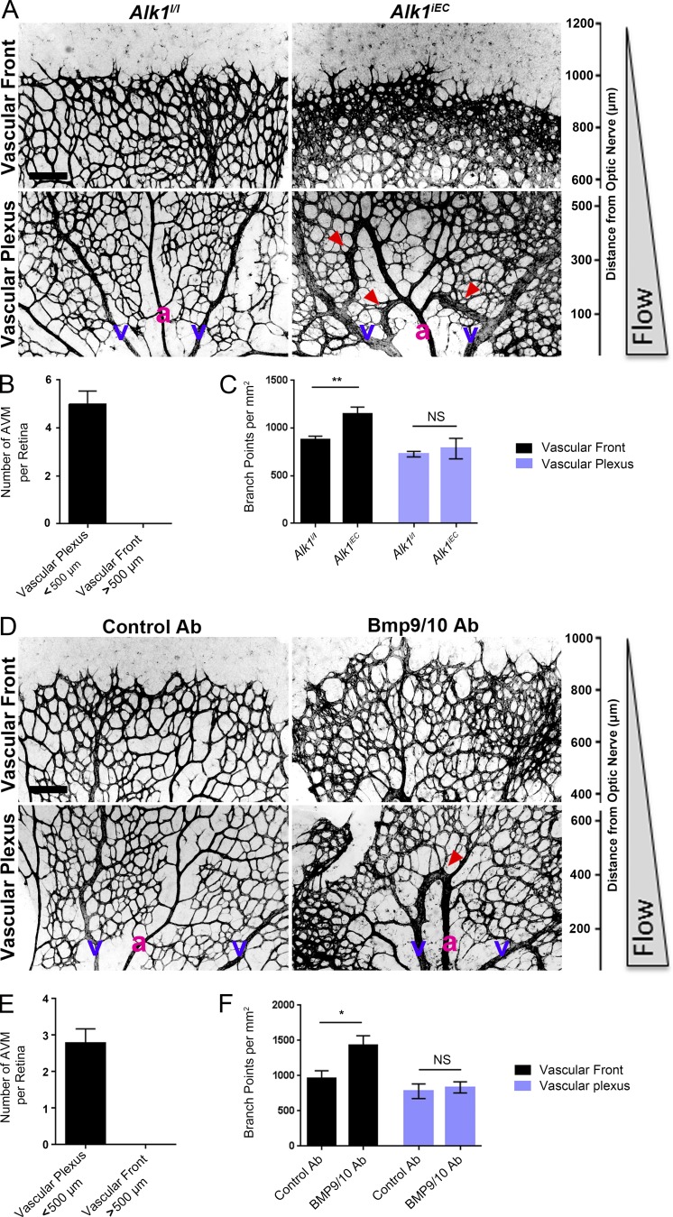
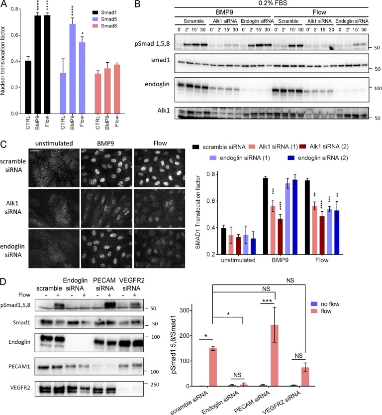
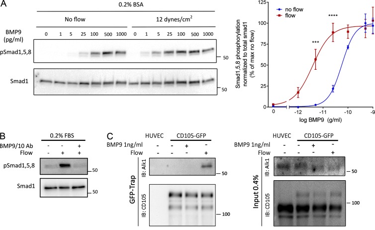
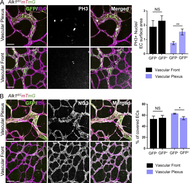
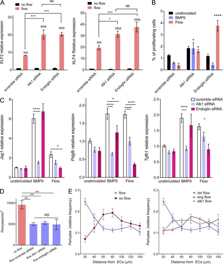
Morphogenesis of the vascular system is strongly modulated by mechanical forces from blood flow. Hereditary hemorrhagic telangiectasia (HHT) is an inherited autosomal-dominant disease in which arteriovenous malformations and telangiectasias accumulate with age. Most cases are linked to heterozygous mutations in Alk1 or Endoglin, receptors for bone morphogenetic proteins (BMPs) 9 and 10. Evidence suggests that a second hit results in clonal expansion of endothelial cells to form lesions with poor mural cell coverage that spontaneously rupture and bleed. We now report that fluid shear stress potentiates BMPs to activate Alk1 signaling, which correlates with enhanced association of Alk1 and endoglin. Alk1 is required for BMP9 and flow responses, whereas endoglin is only required for enhancement by flow. This pathway mediates both inhibition of endothelial proliferation and recruitment of mural cells; thus, its loss blocks flow-induced vascular stabilization. Identification of Alk1 signaling as a convergence point for flow and soluble ligands provides a molecular mechanism for development of HHT lesions.


Figures

Figure 1.
Figure 1.
Effects of blood flow on the development of retinal AVMs under impaired Alk1 signaling. (A) Representative images of P5 retinas stained with isolectin B4 to identify blood vessels in control (Alk1l/l) and Alk1iEC (Cdh5-CreERT2-Alk1iEC) mice after 50 µg tamoxifen injection at P3. (B) Quantification of the number of AVMs in the proximal (<500 µm from the optic nerve) versus distal (>500 µm from the optic nerve) regions of the retina (n = 5). (C) Quantification of the number of branch points in Alk1l/l and Cdh5-CreERT2 (Alk1iEC) mice in the proximal versus distal regions (n = 5, Mann–Whitney; **, P < 0.005). (D) Representative images of P5 retinal vessels from pups injected at P3 with control or BMP9/BMP10 blocking antibodies. v, vein; a, artery. (E and F) Quantification of the number of AVMs (E) or branch points (F) in the proximal and distal regions in mice injected with control or BMP9/BMP10 blocking antibodies (n = 5, Mann–Whitney; *, P < 0.05). Bars, 150 µm. NS, not significant. Error bars represent SEM.
Figure 2.
Figure 2.
Alk1 signaling in response to FSS. (A) Nuclear translocation of Smad1, Smad5, and Smad8 in response to 1 ng/ml BMP9 or 12 dynes/cm2 for 45 min (n = 3–8, ANOVA two-way; *, P < 0.05; ****, P < 0.0001). (B) Representative Western blots of phosphorylated Smad1,5,8, Smad1, Alk1 and endoglin in response to 1 ng/ml BMP9 or 12 dynes/cm2 FSS for the indicated times. Actin was used as a loading control. (C) Representative staining of Smad1 in response to 1 ng/ml BMP9 or 12 dynes/cm2 for 45 min in HUVECs transfected with the indicated siRNAs (siRNA 1: QIAGEN; siRNA 2: GE Healthcare; n = 3–11, two-way ANOVA; **, P < 0.01; ***, P < 0.001; ****, P < 0.0001). Bar, 10 µm. (D) Representative Western blot of Smad1/5/8 phosphorylation in response to 15 min of 12 dynes/cm2 FSS in cells transfected with the indicated siRNAs (n = 3, ANOVA; *, P < 0.05; ***, P < 0.001). NS, not significant.
Figure 3.
Figure 3.
Potentiation of BMP9 responses by FSS. (A) Representative Western blot of Smad1/5/8 phosphorylation in response to the indicated concentrations of BMP9 in serum-free media with or without flow for 45 min (n = 4, two-way ANOVA; ***, P < 0.001; ****, P < 0.0001, BMP9 EC50: 56 pg/ml without flow, 3.6 pg/ml with flow). (B) Representative Western blot of Smad1/5/8 phosphorylation in response to 12 dynes/cm2 for 45 min in the presence or absence of two blocking antibodies against BMP9 and BMP10 (each at 100 ng/ml). (C) Association of endoglin-GFP with endogenous Alk1 in response to 1 ng/ml BMP9 or 12 dynes/cm2 flow for 15 min. GFP-tagged proteins were captured with a GFP-trap and immunoprecipitations analyzed by immunoblotting (IB) as indicated.
Figure 4.
Figure 4.
Effects of Alk1 mosaic deletion on proliferation and pericyte coverage in retinal ECs. (A) Phospho–histone H3 (PH3) staining of P6 retinas of Cdh5-CreERT2 Alk1iEC:mTmG mice injected with low-dose (5 µg) tamoxifen at P3. Unrecombined ECs (GFP−; Alk1 positive) are shown in light purple, and recombined ECs (GFP+; Alk1 negative) are shown in green. The graph shows quantification of PH3-positive cells at the vascular front and vascular plexus in GFP− and GFP+ cells (number of positive nuclei normalized to EC surface area; n = 6 retinas, Mann–Whitney; **, P < 0.005). (B) NG2 staining of P6 retinas after Alk1 mosaic deletion. Graph shows the percentage of EC surface area covered by NG2-positive cells (n = 6 retinas, Mann–Whitney; *, P < 0.05). Bars, 50 µm. NS, not significant.
Figure 5.
Figure 5.
Effect of Alk1 or endoglin depletion on FSS responses. (A) KLF2 and KLF4 message levels by quantitative RT-PCR after flow for 16 h in HUVECs transfected with scrambled, Alk1, or Endoglin siRNA (QIAGEN; n = 3, two-way ANOVA; *, P < 0.05; *** or $$$, P < 0.001; **** or $$$$, P < 0.0001). (B) Endothelial cell proliferation in response to 1 ng/ml BMP9 or 12 dynes/cm2 for 24 h after transfection with scrambled, Alk1, or Endoglin siRNA (QIAGEN). Cell proliferation was measured by incorporation of EdU during the last 4 h. (C) HUVECs were stimulated by 1 ng/ml BMP9 or 12 dynes/cm2 for 3 h. Expression of pericyte recruitment genes (PDGF, TGF-β< and jagged1) was assayed by quantitative RT-PCR (n = 4–12, two-way ANOVA; *, P < 0.05; **, P < 0.01; ****, P < 0.0001). (D) Pericyte density in fibrin gels without stimulation or after 96 h of 10 dynes/cm2 flow on HUVECs transfected with scrambled, Alk1, or Endoglin siRNA (QIAGEN; n = 4–6 gels from four independent experiments, two-way ANOVA; **, P < 0.01). (E) Distribution of pericytes in fibrin gels, relative to the endothelial monolayer with or without 10 dynes/cm2 flow for 96 h. HUVECs were transfected with scrambled, Alk1, or Endoglin siRNA (QIAGEN); n = 5–7 gels from four independent experiments, two-way ANOVA; * or $, P < 0.05; **, P < 0.01; **** or $$$$, P < 0.0001; NS, not significant.

References

    1. Akimoto S., Mitsumata M., Sasaguri T., and Yoshida Y.. 2000. Laminar shear stress inhibits vascular endothelial cell proliferation by inducing cyclin-dependent kinase inhibitor p21(Sdi1/Cip1/Waf1). Circ. Res. 86:185–190. 10.1161/01.RES.86.2.185 - DOI - PubMed
    1. Arthur H.M., Ure J., Smith A.J., Renforth G., Wilson D.I., Torsney E., Charlton R., Parums D.V., Jowett T., Marchuk D.A., et al. 2000. Endoglin, an ancillary TGFbeta receptor, is required for extraembryonic angiogenesis and plays a key role in heart development. Dev. Biol. 217:42–53. 10.1006/dbio.1999.9534 - DOI - PubMed
    1. Aspalter I.M., Gordon E., Dubrac A., Ragab A., Narloch J., Vizán P., Geudens I., Collins R.T., Franco C.A., Abrahams C.L., et al. 2015. Alk1 and Alk5 inhibition by Nrp1 controls vascular sprouting downstream of Notch. Nat. Commun. 6:7264 10.1038/ncomms8264 - DOI - PMC - PubMed
    1. Atri D., Larrivee B., Eichmann A., and Simons M.. 2014. Endothelial signaling and the molecular basis of arteriovenous malformation. Cell. Mol. Life Sci. 71:867–883. 10.1007/s00018-013-1475-1 - DOI - PMC - PubMed
    1. Baeyens N., and Schwartz M.A.. 2016. Biomechanics of vascular mechanosensation and remodeling. Mol. Biol. Cell. 27:7–11. 10.1091/mbc.E14-11-1522 - DOI - PMC - PubMed

Publication types

MeSH terms

LinkOut - more resources