Review
. 2016 Dec;24(12):2043-2053.
doi: 10.1038/mt.2016.182. Epub 2016 Sep 20.

The Mucus Barrier to Inhaled Gene Therapy

Affiliations
Review

The Mucus Barrier to Inhaled Gene Therapy

Gregg A Duncan et al. Mol Ther. 2016 Dec.

Abstract

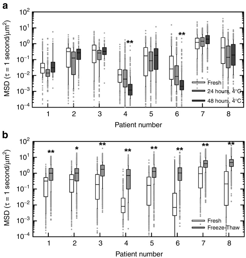
Recent evidence suggests that the airway mucus gel layer may be impermeable to the viral and synthetic gene vectors used in past inhaled gene therapy clinical trials for diseases like cystic fibrosis. These findings support the logic that inhaled gene vectors that are incapable of penetrating the mucus barrier are unlikely to provide meaningful benefit to patients. In this review, we discuss the biochemical and biophysical features of mucus that contribute its barrier function, and how these barrier properties may be reinforced in patients with lung disease. We next review biophysical techniques used to assess the potential ability of gene vectors to penetrate airway mucus. Finally, we provide new data suggesting that fresh human airway mucus should be used to test the penetration rates of gene vectors. The physiological barrier properties of spontaneously expectorated CF sputum remained intact up to 24 hours after collection when refrigerated at 4 °C. Conversely, the barrier properties were significantly altered after freezing and thawing of sputum samples. Gene vectors capable of overcoming the airway mucus barrier hold promise as a means to provide the widespread gene transfer throughout the airway epithelium required to achieve meaningful patient outcomes in inhaled gene therapy clinical trials.


Figures

Figure 1
Figure 1
Mucus in the airways of humans without lung disease. (a) Histological staining of human primary bronchial epithelial cell cultures showing the airway surface liquid (ASL) composed of the periciliary layer (PCL) and the airway mucus gel layer (mucus). Reproduced with permission from. (b) Scanning electron micrograph of human airway mucus collected from an individual without lung disease (Scale bar = 500 nm). Reproduced with permission from. (c) Schematic of mucin subunits connected via disulfide bonds between cysteine domains to form the airway mucus gel. Reproduced with permission from.
Figure 2
Figure 2
Mucus in the airways of humans with obstructive lung diseases. (a) Confocal microscopy images of human bronchial epithelial-derived mucus hydrogels at 1.5 and 2.5% total mucus solid content (Scale bars = 500 µm). Mucins were fluorescently labeled with rhodamine-conjugated wheat germ agglutinin. Reproduced with permission from. (b) Scanning electron micrographs of cystic fibrosis (CF) airway mucus before (native) and after reduction of disulfide cross-links between mucin fibers by N-acetyl cysteine (NAC) treatment (Scale bar = 300 nm). Reproduced with permission from. (c) Confocal microscopy images of CF airway mucus composed of mucin (red; anti-MUC5AC/MUC5B) and DNA (green; YO-PRO I Iodide) (Scale bar = 20 µm). Reproduced with permission from.
Figure 3
Figure 3
Adhesive and steric trapping of nanoparticles and gene vectors in airway mucus. (a) Representative 20-second trajectories of 200 nm nonmucoadhesive, PEG-coated polystyrene nanoparticles (PS-PEG NP), adenovirus (AdV), and adeno-associated virus serotype 5 (AAV5) in cystic fibrosis (CF) airway mucus, as captured using multiple particle tracking (MPT). Reproduced with permission from. (b) Representative 3-second trajectories of 100, 200, and 500 nm PS-PEG NP in human airway mucus from individuals without lung disease, as captured using MPT. Reproduced with permission from.
Figure 4
Figure 4
Biophysical techniques used to assess the barrier properties of airway mucus. (a) Schematic of a diffusion chamber experiment showing nanoparticle (NP) diffusion from the donor compartment, across a mucus layer, and into the acceptor compartment. NP concentrations in the donor and acceptor compartments are measured to assess the percentage of gene vectors that penetrate a mucus layer with a designated thickness. (b) Schematic of fluorescence recovery after photobleaching (FRAP) experiments showing recovery of fluorescence into a rapidly photo-bleached region due to the diffusion of unbleached gene vectors through mucus. The time to 50% fluorescence recovery (τ1/2) in the bleached region is measured to assess gene vector diffusion rate. The mobile and immobile fraction is determined based on the fraction of fluorescence recovery compared with the prebleached fluorescence intensity. (c) Schematic of particle tracking experiments showing NP trajectories based on their tracked motion within mucus. Using these trajectories, the mean squared displacement (MSD) at designated timescale (τ) is determined for up to thousands of individual gene vectors.
Figure 5
Figure 5
Effects of storage on cystic fibrosis (CF) sputum microstructural properties. (a–b) Box-and-whisker plots of measured mean squared displacement (MSD) in µm2 at time scale τ = 1 second of 100 nm PEG-coated polystyrene nanoparticles (PS-PEG NP) in spontaneously expectorated sputum samples from eight CF patients. (a) Transport rates of 100 nm PS-PEG NP in CF sputum samples immediately after collection (Fresh; white bars), after 24 hours storage at 4ºC (24 hours, 4ºC; light gray bar) and after 48 hours storage at 4ºC (48 hours, 4ºC; dark gray bar). (b) Transport rates of 100 nm PS-PEG NP in CF sputum samples immediately after collection (fresh; white bars) and after being frozen at −80ºC overnight and subsequently thawed on ice (freeze-thaw; light gray bar). A Mann-Whitney test was used to determine statistically significant differences in MSD values (*P < 0.05; **P < 0.01). The data presented in parts a and b were collected from two independent patient cohorts. To avoid a concern of potential intrasample variation, identical aliquots of individual samples were used to compare MSD of 100 nm PS-PEG NP in fresh mucus versus mucus stored at different conditions. Briefly, 0.5 µl solution of fluorescently labeled 100 nm PS-PEG NP was added to 30 µl of CF sputum in a custom-made microwell. Samples were imaged at room temperature using an Axio Observer inverted epifluorescence microscope equipped with 100x/1.46 NA oil-immersion objective. Movies were recorded over 20 seconds at an exposure time of 67 milliseconds (i.e., 15 frames per second) by an Evolve 512 EMCCD camera. Movies were analyzed using a custom-made MATLAB code to simultaneously extract x, y-coordinates of >500 NP per sample aliquot to calculate MSD values. One 30 µl aliquot of CF sputum from each patient was assessed following sample collection (i.e., fresh) and after storage at 4ºC for 24 and 48 hours. A second 30 µl aliquot was assessed following sample collection and after freezing at −80ºC overnight and thawing on ice. For evaluating the effect of freeze-thaw on CF sputum barrier properties, fresh yellow-green (505/515 nm) fluorescent 100 nm PS-PEG NP were added to the freeze-thawed aliquot due to concerns over of the effects of freezing on the red (580/605 nm) fluorescent 100 nm PS-PEG NP previously added to assess the fresh sample. To confirm the particle sets were comparable, the size and ζ-potential for each set of 100 nm PS-PEG were measured by dynamic light scattering and laser Doppler anemometry, respectively. Yellow-green and red PS-PEG NP had diameters of 104 ± 0.3 and 107 ± 1.3 nm; and ζ-potential of −4.4 ± 0.3 and −4.7 ± 0.2 mV, respectively.

References

    1. Griesenbach, U and Alton, EW; UK Cystic Fibrosis Gene Therapy Consortium (2009). Gene transfer to the lung: lessons learned from more than 2 decades of CF gene therapy. Adv Drug Deliv Rev 61: 128–139. - PubMed
    1. Ferrari, S, Geddes, DM and Alton, EW (2002). Barriers to and new approaches for gene therapy and gene delivery in cystic fibrosis. Adv Drug Deliv Rev 54: 1373–1393. - PMC - PubMed
    1. Sanders, N, Rudolph, C, Braeckmans, K, De Smedt, SC and Demeester, J (2009). Extracellular barriers in respiratory gene therapy. Adv Drug Deliv Rev 61: 115–127. - PMC - PubMed
    1. Lai, SK, Wang, YY and Hanes, J (2009). Mucus-penetrating nanoparticles for drug and gene delivery to mucosal tissues. Adv Drug Deliv Rev 61: 158–171. - PMC - PubMed
    1. Cone, RA (2009). Barrier properties of mucus. Adv Drug Deliv Rev 61: 75–85. - PubMed