. 2016 Sep 20:6:33488.
doi: 10.1038/srep33488.

Multi-Phenotypic subtyping of circulating tumor cells using sequential fluorescent quenching and restaining

Affiliations

Multi-Phenotypic subtyping of circulating tumor cells using sequential fluorescent quenching and restaining

Daniel L Adams et al. Sci Rep. .

Abstract

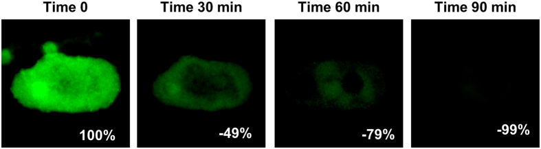
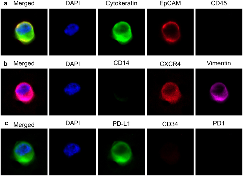
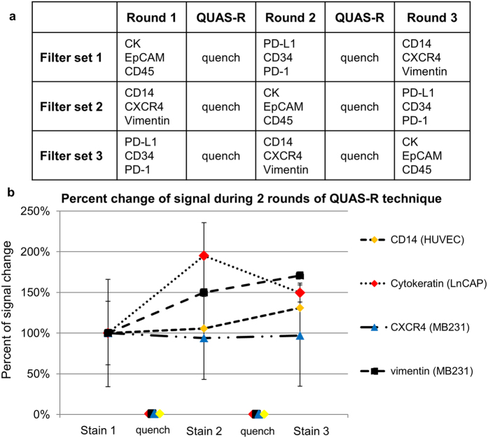
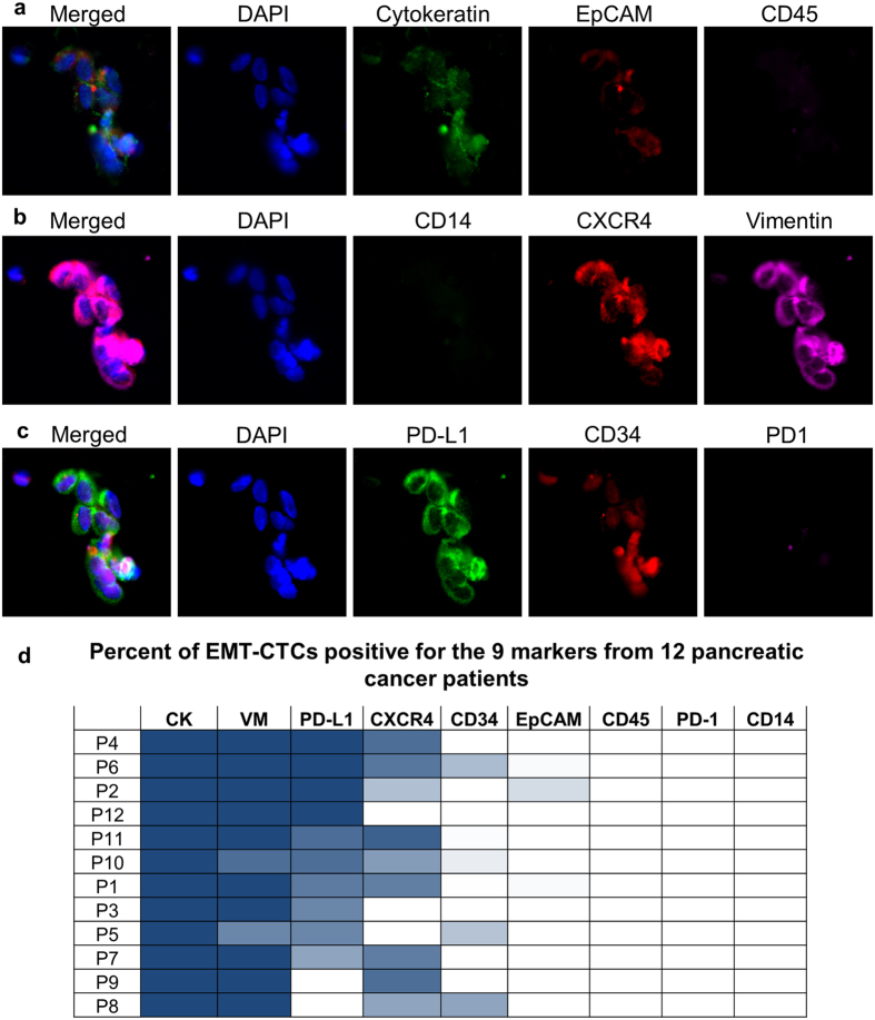
In tissue biopsies formalin fixed paraffin embedded cancer blocks are micro-sectioned producing multiple semi-identical specimens which are analyzed and subtyped proteomically, and genomically, with numerous biomarkers. In blood based biopsies (BBBs), blood is purified for circulating tumor cells (CTCs) and clinical utility is typically limited to cell enumeration, as only 2-3 positive fluorescent markers and 1 negative marker can be used. As such, increasing the number of subtyping biomarkers on each individual CTC could dramatically enhance the clinical utility of BBBs, allowing in depth interrogation of clinically relevant CTCs. We describe a simple and inexpensive method for quenching the specific fluors of fluorescently stained CTCs followed by sequential restaining with additional biomarkers. As proof of principle a CTC panel, immunosuppression panel and stem cell panel were used to sequentially subtype individual fluorescently stained patient CTCs, suggesting a simple and universal technique to analyze multiple clinically applicable immunomarkers from BBBs.


Conflict of interest statement

D. Adams and CM. Tang are employees at Creatv Microtech, Inc. S. Stefansson is an employee of HeMemics Biotechnologies, Inc.

Figures

Figure 1
Figure 1. Tracking the quench time of borohydride solution in the removal of visible and quantifiable fluorescence from patient blood cells.
A Cytokeratin positive cell isolated from a pancreatic cancer patient was identified, imaged and the signal measured at time 0. Borohydride solution was added and the cell was imaged 30 min, 60 min and 90 after induction of borohydride. After 90 min, 99% of the original fluorescence had been quenched. Box scale = 45 μm.
Figure 2
Figure 2. Visual example of a MDA-MB-231 cell through the 2 QUAS-R rounds.
(a) After filtration, a MDA-MB-231 cell was stained with the CTC stain Cytokeratin, EpCAM and CD45. (b) The cell was quenched by QUAS-R and stained with CD14, CXCR4 and Vimentin. (c) The cell was quenched again by QUAS-R and stained with PD-L1, CD34, and PD1. Box scale = 45.
Figure 3
Figure 3. Overview of experimental design and representative examples of the percent change of signal intensity when a marker stain was used on the first round, second round or third round of staining.
(a) Stains were tested on 5 cell lines at three separate time points. A 1st set of stains were used followed by quenching. After quench, a 2nd set of stains were used followed by quenching. After quenching, a 3rd set of stains were used. After each round, cells were imaged and quantified. b. Representative of signal intensities shows no degradation irregardless of whether the stains were used first, second or third. A repeated measure ANOVA found no significant difference in signal intensity between the 3 staining times [vimentin MDA-MB-231 (p = 0.201), cytokeratin LNCaP (p = 0.291), CD14 HUVEC (p = 0.499), and CXCR4 MDA-MB-231 (p = 0.857)]. Full comparison of all cells and all markers is found in Supplementary Figures 2 and 10.
Figure 4
Figure 4. QUAS-R on patient derived EMT-like CTCs for subtyping cells and screening drug targets.
(a) A proof of principle study of 12 pancreatic cancer patient samples were imaged with the CTC stains CK, EpCAM and CD45. EMT-Like CTCs were enumerated and imaged. (b) QUAS-R was performed and cells were stained with the subtyping markers CD14, CXCR4, and Vimentin. (c) QUAS-R was then performed again and cells stained for the drug targets PD-L1, CD34 and PD1. (d) A total of 764 EMTCTCs with a median of 10 cells per sample were measured for presence of the 9 markers. Presence of each marker in each patient is represented as a heat map of percent EMT-CTCs positive for each stain. VM = vimentin, CK = cytokeratin, numerical values can be found in Supplementary Figure 11. Box scale = 75 μm.

Similar articles

Cited by

References

    1. Adams D. L. et al. Precision microfilters as an all in one system for multiplex analysis of circulating tumor cells. RSC Advances 6, 6405–6414, doi: 10.1039/C5RA21524B (2016). - DOI - PMC - PubMed
    1. Adams D. L. et al. Cytometric characterization of circulating tumor cells captured by microfiltration and their correlation to the cellsearch((R)) CTC test. Cytometry. Part A: the journal of the International Society for Analytical Cytology 87, 137–144, doi: 10.1002/cyto.a.22613 (2015). - DOI - PubMed
    1. Adams D. L. et al. The systematic study of circulating tumor cell isolation using lithographic microfilters. RSC Advances 4, 4334–4342, doi: 10.1039/C3RA46839A (2014). - DOI - PMC - PubMed
    1. Alix-Panabieres C. & Pantel K. Challenges in circulating tumour cell research. Nature reviews. Cancer 14, 623–631, doi: 10.1038/nrc3820 (2014). - DOI - PubMed
    1. Allard W. J. et al. Tumor cells circulate in the peripheral blood of all major carcinomas but not in healthy subjects or patients with nonmalignant diseases. Clin Cancer Res 10, 6897–6904, doi: 10.1158/1078-0432.CCR-04-0378 (2004). - DOI - PubMed

Publication types

MeSH terms