Review
. 2016 Oct;57(3):R127-42.
doi: 10.1530/JME-16-0124.

Disorders of the calcium-sensing receptor and partner proteins: insights into the molecular basis of calcium homeostasis

Affiliations
Free PMC article
Review

Disorders of the calcium-sensing receptor and partner proteins: insights into the molecular basis of calcium homeostasis

Fadil M Hannan et al. J Mol Endocrinol. 2016 Oct.
Free PMC article

Abstract

The extracellular calcium (Ca(2+) o)-sensing receptor (CaSR) is a family C G protein-coupled receptor, which detects alterations in Ca(2+) o concentrations and modulates parathyroid hormone secretion and urinary calcium excretion. The central role of the CaSR in Ca(2+) o homeostasis has been highlighted by the identification of mutations affecting the CASR gene on chromosome 3q21.1. Loss-of-function CASR mutations cause familial hypocalciuric hypercalcaemia (FHH), whereas gain-of-function mutations lead to autosomal dominant hypocalcaemia (ADH). However, CASR mutations are only detected in ≤70% of FHH and ADH cases, referred to as FHH type 1 and ADH type 1, respectively, and studies in other FHH and ADH kindreds have revealed these disorders to be genetically heterogeneous. Thus, loss- and gain-of-function mutations of the GNA11 gene on chromosome 19p13.3, which encodes the G-protein α-11 (Gα11) subunit, lead to FHH type 2 and ADH type 2, respectively; whilst loss-of-function mutations of AP2S1 on chromosome 19q13.3, which encodes the adaptor-related protein complex 2 sigma (AP2σ) subunit, cause FHH type 3. These studies have demonstrated Gα11 to be a key mediator of downstream CaSR signal transduction, and also revealed a role for AP2σ, which is involved in clathrin-mediated endocytosis, in CaSR signalling and trafficking. Moreover, FHH type 3 has been demonstrated to represent a more severe FHH variant that may lead to symptomatic hypercalcaemia, low bone mineral density and cognitive dysfunction. In addition, calcimimetic and calcilytic drugs, which are positive and negative CaSR allosteric modulators, respectively, have been shown to be of potential benefit for these FHH and ADH disorders.

Keywords: G protein; kidney; parathyroid and bone; signal transduction.


Figures

Figure 1
Figure 1
Overview of Ca2+o homeostasis. The parathyroid CaSR senses reductions in Ca2+o, which leads to a rapid rise in PTH secretion. The increased circulating PTH acts via the PTH1-receptor (PTH1R) in the kidneys and bone. The skeletal effects of PTH are to increase bone resorption, thereby releasing calcium into the extracellular fluid. In the kidney, PTH increases calcium reabsorption and stimulates the proximal renal tubular 1-α-hydroxylase (1αOHase) enzyme, which promotes the synthesis of the active 1,25-dihydroxyvitamin D3 (1,25D3) metabolite from 25-hydroxyvitamin D3 (25D3), which is the major circulating form of vitamin D. The elevated 1,25D3 acts on the intestine via the vitamin D receptor (VDR) to increase the absorption of dietary calcium. Thus, in response to hypocalcaemia, the secretion of PTH, by these direct and indirect actions leads to the restoration of normocalcaemia. The kidney CaSR senses reductions in Ca2+o and promotes urinary calcium reabsorption independent of the actions of PTH. The rise in Ca2+o and 1,25D3 concentrations mediated by PTH act on the parathyroid glands to induce feedback inhibition of further PTH secretion.
Figure 2
Figure 2
Role of the CaSR, Gα11 and AP2 complex in the regulation of PTH secretion and renal tubular calcium reabsorption. The binding of calcium (red filled-in circle, Ca) to the extracellular bilobed venus fly trap (VFT) domain of the CaSR (grey) results in Gα11 (yellow)-dependent stimulation of phospholipase C-β (PLCβ) (dark blue) activity, which catalyses the formation of inositol 1,4,5-trisphosphate (IP3) and diacylglycerol (DAG) from phosphatidylinositol 4,5-bisphosphate (PIP2). An accumulation of IP3 mediates the rapid release of calcium into the cytosol from intracellular stores, whereas DAG activates the MAPK cascade. These intracellular signalling events lead to a decrease in PTH secretion from the parathyroid chief cell and reduction in renal tubular calcium reabsorption. CaSR cellsurface expression is regulated by agonist-driven insertional signalling (ADIS) (not shown) (Grant et al. 2011) and also by an endocytic complex comprising clathrin, β-arrestin (green) and the AP2 complex (orange), which traffic this GPCR to the endosomal-lysosomal degradation pathway (light blue) or recycle the CaSR back to the cell surface (Breitwieser 2013). Loss- and gain-of-function mutations of the CaSR lead to FHH1 and ADH1, respectively, whereas loss- and gain-of function mutations of the Gα11-subunit are associated with FHH2 and ADH2, respectively. Loss-offunction mutations of the AP2σ-subunit lead to FHH3.
Figure 3
Figure 3
(A) Schematic representation of the genomic organisation of the CASR gene showing mutational hotspots for disease-associated missense mutations. The CASR gene consists of six coding exons (2–7), and the start (ATG) and stop (TGA) codons are in exons 2 and 7, respectively. The 5′ portion of exon 2 and the 3′ portion of exon 7 are untranslated (open boxes). The 3′ portion of exon 2, exons 3, 4, 5 and 6 and the 5′ portion of exon 7, encode the extracellular domain (light grey). The mid portion of exon 7 encodes the transmembrane (dark grey) and intracellular (black) domains. More than 170 different missense CASR mutations have been reported in patients with FHH1, NSHPT, adult-onset PHPT, ADH1 and Bartter syndrome type V (Hannan & Thakker 2013). These mutations affect >110 different codons scattered throughout the CASR gene. Twenty-eight codons (>20% of all mutated codons) represent mutational hotspots as they are the site of recurrent missense mutations (solid lines) that have been reported in three or more probands (mutational frequency of >1.5%), and/or affected by multiple (3 or more) different missense mutations (dashed lines). Mutations affecting these codons are clustered in three regions, which are the 2nd peptide loop of the extracellular domain, venus flytrap (VFT) cleft region Ca2+o binding site, and the region encompassing transmembrane domains (TMD) 6 and 7. Codons 173, 221, 297 and 802 (thickened lines) are the site of missense mutations that may lead to a loss- or gain-of-function and are termed ‘switch’ codons or residues. (B) Homology modelling of the CaSR VFT cleft region ligand-bound Ca2+o binding site based on the crystal structure of the metabotropic glutamate receptor type 1. The Leu173 (L173) and Pro221 (P221) switch residues are predicted to be located in short α-helices within lobes 1 and 2, respectively, that form the entrance to the Ca2+o binding site within the VFT cleft. The L173 and P221 side chains are shown in purple, the side chains of predicted Ca2+o binding residues (Ser147 (S147), Ser170 (S170), Asp190 (D190), Tyr218 (Y218) and Glu297 (E297)) are shown in cyan, and a bound calcium ion is shown as a green sphere. The side chains of L173 and P221 are predicted to extend across the entrance to the Ca2+o binding site. Mutations affecting these residues may lead to opposing effects on CaSR function by influencing the entry and binding of calcium within the VFT cleft region. Adapted, with permission, from Hannan FM, Nesbit MA, Zhang C, Cranston T, Curley AJ, Harding B, Fratter C, Rust N, Christie PT, Turner JJ, et al. (2012) Identification of 70 calcium-sensing receptor mutations in hyper- and hypo-calcaemic patients: evidence for clustering of extracellular domain mutations at calcium-binding sites. Human Molecular Genetics 21 2768–2778. Copyright 2012 Oxford University Press.
Figure 4
Figure 4
(A) Schematic representation of the genomic organisation of the GNA11 gene showing germline disease-associated mutations. The GNA11 gene consists of 7 coding exons (1–7), and the start (ATG) and stop (TGA) codon are in exons 1 and 7, respectively. The 5′ portion of exon 1 and the 3′ portion of exon 7 are untranslated (open boxes). The 3′ portion of exon 2, exon 3 and 5′ portion of exon 4 encode the Gα11 helical domain, which is connected by two short peptides, termed linker 1 (L1) and linker 2 (L2), to the GTPase domain. The Gα11 GTPase domain is encoded by the 3′ portion of exon 1, 5′ portion of exon 2, 3′ portion of exon 4 and exons 5–7. Three flexible regions, termed switch regions 1–3 (S1–S3, shown in blue), which undergo conformational changes during Gα11 activation are encoded by exons 4 and 5. The location of reported FHH2- and ADH2-causing mutations is shown. (B) Three-dimensional homology model of Gα11 showing the location of residues mutated in FHH2 (blue) and ADH2 (red). The homology model is based on the crystal structure of Gαq (PDB accession number 3AH8) (Nishimura et al. 2010), which shares 90% amino acid identity with Gα11. The Gα11 helical and GTPase domains are shown bound to GDP aluminium fluoride (GDP-AlF4, green), which is a non-hydrolysable analogue of GTP. The three flexible switch regions are highlighted in cyan, and the L1 and L2 peptides are shown in yellow. The β2–β3 hairpin loop, which comprises part of the Gα–GPCR interface, is shown in orange. Adapted, with permission, from Nesbit MA, Hannan FM, Howles SA, Babinsky VN, Head RA, Cranston T, Rust N, Hobbs MR, Heath H 3rd & Thakker RV (2013) Mutations affecting G-protein subunit alpha11 in hypercalcemia and hypocalcemia. New England Journal of Medicine 368 2476–2486. Copyright 2013 Massachusetts Medical Society.
Figure 5
Figure 5
Three-dimensional model of Gαs (brown) bound to the β2-adrenergic receptor (β2AR, green) showing location of residues homologous to the Gα11 Ile199 and Phe341 residues, which are mutated in FHH2 and ADH2, respectively. Gαs residues homologous to the Gα11 Ile199 and Phe341 residues (red) are located within a hydrophobic region at the GPCR–Gα interface (black open circle). The β2AR–Gαs interface is formed by residues located within the β1 strand, hairpin loop linking the β2 and β3 strands, and the α5 helix of the Gαs protein, which interact with intracellular loop 2 (IL2) of the β2AR. The Gαs Val217 (V217) and Phe376 (F376) residues, which are homologous to the Gα11 Ile199 and Phe341 residues, comprise part of a hydrophobic pocket (curved line) at the Gα-subunit surface, which facilitates the docking of the β2AR IL2 with the Gαs protein (Rasmussen et al. 2011).
Figure 6
Figure 6
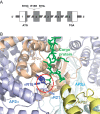
(A) Schematic representation of the genomic organisation of the AP2S1 gene showing the location of FHH3-causing mutations. The AP2S1 gene consists of 5 coding exons (1–5), and the start (ATG) and stop (TGA) codon are in exons 1 and 5, respectively. The 5′ portion of exon 1 and the 3′ portion of exon 5 are untranslated (open boxes). The AP2σ protein is encoded by the 3′ portion of exon 1, exons 2, 3, 4, and the 5′ portion of exon 5 (dark grey). The FHH3-causing mutations all affect the Arg15 residue and comprise Arg15Cys (R15C), Arg15His (R15H) and Arg15Leu (R15L) missense substitutions. (B) Three-dimensional model of the heterotetrameric AP2 complex, which comprises α- (purple), β- (yellow), μ- (light blue) and σ- (light brown) subunits (PDB accession number 2JKR, (Kelly et al. 2008)). The AP2 complex is bound to a cargo protein recognition motif (green) via key polar contacts (shown in the red dashed circle) involving the AP2σ Arg15 (R15) residue (dark blue) and Arg21 (R21) residue of the AP2α-subunit. Adapted from Nesbit MA et al. Nat Genet. 2013 45:93–97. Adapted, with permission, from Nesbit MA, Hannan FM, Howles SA, Reed AA, Cranston T, Thakker CE, Gregory L, Rimmer AJ, Rust N, Graham U, et al. (2013) Mutations in AP2S1 cause familial hypocalciuric hypercalcemia type 3. Nature Genetics 45 93–97.

References

    1. Atay Z, Bereket A, Haliloglu B, Abali S, Ozdogan T, Altuncu E, Canaff L, Vilaca T, Wong BY, Cole DE, et al. 2014. Novel homozygous inactivating mutation of the calcium-sensing receptor gene (CASR) in neonatal severe hyperparathyroidism-lack of effect of cinacalcet. Bone 64 102–107. (10.1016/j.bone.2014.04.010) - DOI - PubMed
    1. Babinsky VN, Hannan FM, Gorvin CM, Howles SA, Nesbit MA, Rust N, Hanyaloglu AC, Hu J, Spiegel AM, Thakker RV. 2016. Allosteric modulation of the calcium-sensing receptor rectifies signaling abnormalities associated with G-protein alpha-11 mutations causing hypercalcemic and hypocalcemic disorders. Journal of Biological Chemistry 291 10876–10885. (10.1074/jbc.M115.696401) - DOI - PMC - PubMed
    1. Bai M, Quinn S, Trivedi S, Kifor O, Pearce SH, Pollak MR, Krapcho K, Hebert SC, Brown EM. 1996. Expression and characterization of inactivating and activating mutations in the human Ca2+o-sensing receptor. Journal of Biological Chemistry 271 19537–19545. (10.1074/jbc.271.32.19537) - DOI - PubMed
    1. Breitwieser GE. 2013. The calcium sensing receptor life cycle: trafficking, cell surface expression, and degradation. Best Practice & Research. Clinical Endocrinology & Metabolism 27 303–313. (10.1016/j.beem.2013.03.003) - DOI - PubMed
    1. Breitwieser GE, Gama L. 2001. Calcium-sensing receptor activation induces intracellular calcium oscillations. American Journal of Physiology: Cell Physiology 280 C1412–1421. - PubMed

Publication types

MeSH terms

LinkOut - more resources