. 2016 Nov 18;84(12):3423-3433.
doi: 10.1128/IAI.00496-16. Print 2016 Dec.

Filifactor alocis Promotes Neutrophil Degranulation and Chemotactic Activity

Affiliations

Filifactor alocis Promotes Neutrophil Degranulation and Chemotactic Activity

Cortney L Armstrong et al. Infect Immun. .

Abstract

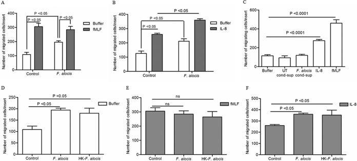
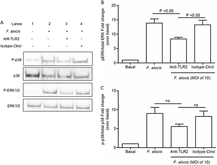
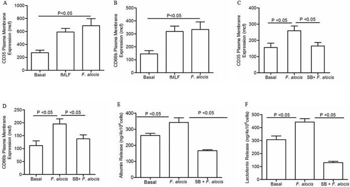
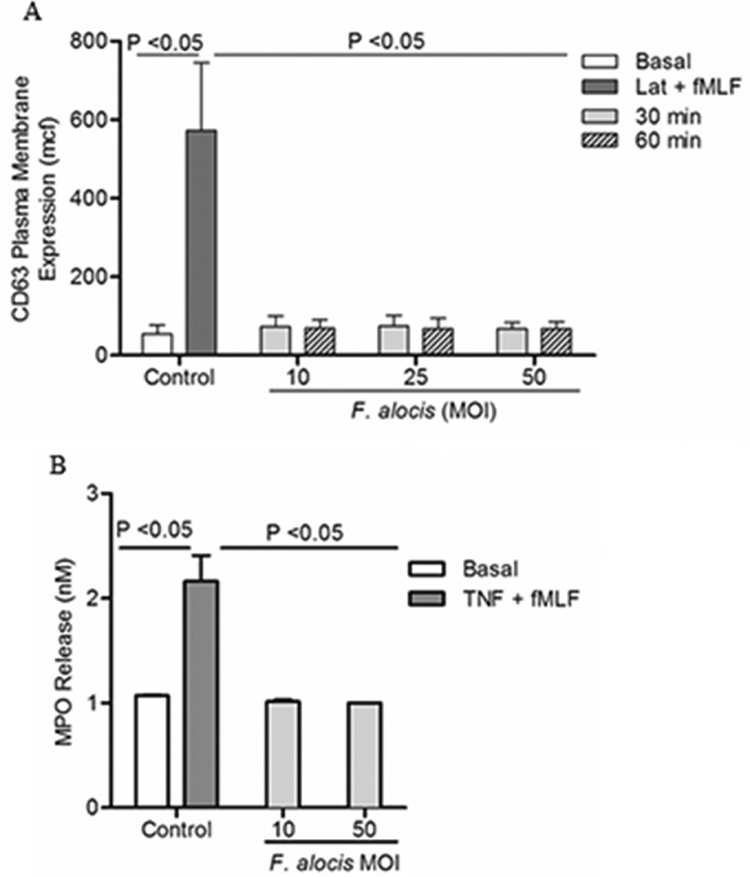
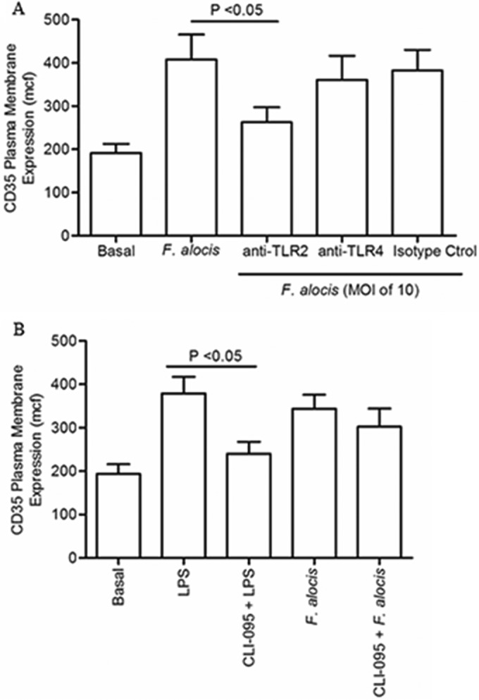
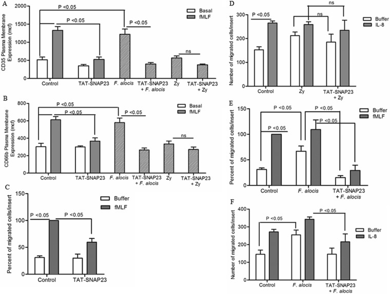
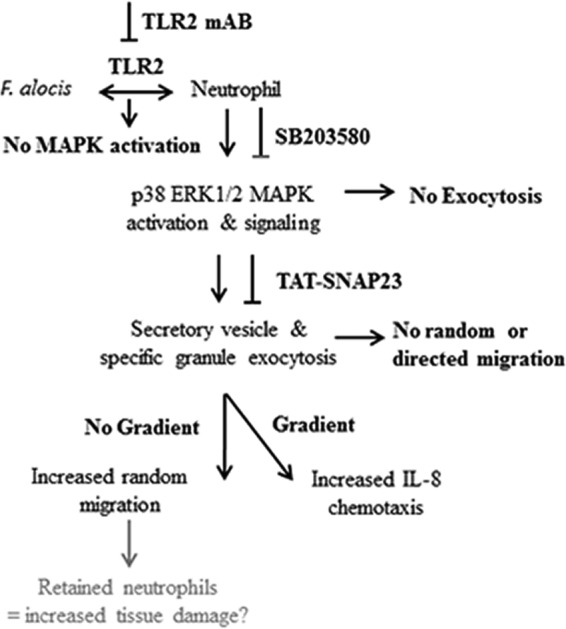
Filifactor alocis is a recently recognized periodontal pathogen; however, little is known regarding its interactions with the immune system. As the first-responder phagocytic cells, neutrophils are recruited in large numbers to the periodontal pocket, where they play a crucial role in the innate defense of the periodontium. Thus, in order to colonize, successful periodontal pathogens must devise means to interfere with neutrophil chemotaxis and activation. In this study, we assessed major neutrophil functions, including degranulation and cell migration, associated with the p38 mitogen-activated protein kinase (MAPK) signaling pathway upon challenge with F. alocis. Under conditions lacking a chemotactic gradient, F. alocis-challenged neutrophils had increased migration compared to uninfected cells, indicating that F. alocis increases chemokinesis in human neutrophils. In addition, neutrophil chemotaxis induced by interleukin-8 was significantly enhanced when cells were challenged with F. alocis, compared to noninfected cells. Similar to live bacteria, heat-killed F. alocis induced both random and directed migration of human neutrophils. The interaction of F. alocis with Toll-like receptor 2 induced granule exocytosis along with a transient ERK1/2 and sustained p38 MAPK activation. Moreover, F. alocis-induced secretory vesicle and specific granule exocytosis were p38 MAPK dependent. Blocking neutrophil degranulation with TAT-SNAP23 fusion protein significantly reduced the chemotactic and random migration induced by F. alocis Therefore, we propose that induction of random migration by F. alocis will prolong neutrophil traffic time in the gingival tissue, and subsequent degranulation will contribute to tissue damage.


Figures

FIG 1
FIG 1
Effect of F. alocis stimulation on neutrophil chemotaxis. Neutrophils were left unchallenged (control), challenged with F. alocis (30 min), or challenged with heat-killed F. alocis (HK-F. alocis; 30 min). (A to F) Following the bacterial challenge, cells were placed in the upper chamber of the transwell system, and after 30 min of incubation, the membrane was stained with a HEMA 3 stain set kit. Chemotaxis was assessed by light microscopic examination (magnification, ×100). (A) Buffer or fMLF (100 nM) was placed in the lower well. Data are expressed as mean numbers ± standard errors of the mean (SEM) of migrated cells/insert from 9 independent experiments. (B) Buffer or IL-8 (100 ng/ml) was placed in the lower well. Data are expressed as mean numbers ± SEM of migrated cells/insert from 5 independent experiments. (C) Unstimulated cells were placed in the upper chamber of the transwell plate, and buffer, conditioned supernatant collected from unstimulated cells (UT-cond-sup), or conditioned supernatant collected after 60 min of stimulation with F. alocis (F. alocis-cond-sup), IL-8 (100 ng/ml), or fMLF (100 nM) was placed in the lower well. Data are means ± SEM from 6 independent experiments. (D to F) Buffer (D), fMLF (E), or IL-8 (F) was placed in the lower well. Data are expressed as mean numbers ± SEM of migrated cells/insert from 5 independent experiments.
FIG 2
FIG 2
F. alocis-induced ERK1/2 and p38 MAPK activation in human neutrophils. Neutrophils were unchallenged (basal), stimulated with fMLF (300 nM, 1 min), or challenged with F. alocis for the indicated times. Cells were lysed, and proteins were separated by SDS-PAGE and immunoblotted for phospho-p38 (P-p38) or phospho-ERK1/2 (P-ERK1/2). Blots were stripped and reblotted for total p38 (p38) or total ERK1/2 (ERK1/2), respectively. (A) Representative immunoblot of 5 independent experiments. (B) Densitometric analysis of the 5 immunoblots for P-p38 or P-ERK1/2 normalized to the total amount of p38 or ERK1/2, respectively. Data are expressed as the mean ratio ± SEM of phosphorylated to total kinase.
FIG 3
FIG 3
TLR2 activation is involved in F. alocis-induced phosphorylation of both ERK1/2 and p38 MAPK. Neutrophils were unchallenged (basal), challenged with F. alocis (MOI of 10, 15 min), or pretreated with either anti-TLR2 MAb or isotype control (isotype-Ctrol), followed by F. alocis challenge. Cells were lysed and proteins separated by SDS-PAGE and immunoblotted for phospho-p38 (P-p38) or phospho-ERK1/2 (P-ERK1/2). Blots were stripped and reblotted for total p38 (p38) or total ERK1/2 (ERK1/2), respectively. (A) Representative immunoblot of 4 independent experiments. (B) Densitometric analysis of the 4 immunoblots for P-ERK1/2/total ERK1/2. (C) Densitometric analysis of the 4 immunoblots for P-p38 MAPK/total p38 MAPK. Data are expressed as mean fold changes ± SEM over the basal level of the phosphorylated/total kinase ratio.
FIG 4
FIG 4
F. alocis stimulation of secretory vesicle and specific granule exocytosis is p38 MAPK dependent. Neutrophils were left unchallenged (basal), challenged with fMLF (300 nM, 5 min), challenged with F. alocis (MOI of 10, 30 min), or pretreated for 30 min with SB203580 followed by F. alocis challenge (SB + F. alocis). (A to D) Secretory vesicle and specific granule exocytosis were determined by the increase in plasma membrane expression of the CD35 or CD66b marker, respectively, by flow cytometry. Data are expressed as the mean channel of fluorescence (mcf) ± SEM from 5 independent experiments. (E and F) Supernatants from all of the different experimental conditions were collected, and the release of albumin or lactoferrin to determine secretory vesicle or specific granule exocytosis, respectively, was measured by ELISA. Data from albumin or lactoferrin release are expressed as means ± SEM in ng/4 × 106 cells from 5 independent experiments for albumin and 6 independent experiments for lactoferrin.
FIG 5
FIG 5
F. alocis challenge does not induce azurophil granule exocytosis. Neutrophils were left unchallenged (basal), were pretreated with latrunculin-A (1 μM, 30 min) followed by fMLF stimulation (Lat + fMLF, 300 nM, 5 min), challenged with TNF (2 ng/ml, 10 min) followed by fMLF stimulation (TNF + fMLF, 300 nM, 5 min), or challenged with F. alocis (MOI of 10, 25, and 50 for 30 or 60 min). (A) Azurophil granule exocytosis was determined by the increase in plasma membrane expression of CD63 by flow cytometry. Data are expressed as mean mcf ± SEM from 3 independent experiments. (B) Extracellular release of myeloperoxidase (MPO), to determine azurophil granule exocytosis, was measured as described in Materials and Methods. Data from MPO release are expressed as means ± SEM in nM from 3 independent experiments.
FIG 6
FIG 6
F. alocis interaction with TLR2 triggered secretory vesicle exocytosis. (A) Neutrophils were left unchallenged (basal), challenged with F. alocis (M0I of 10, 30 min), or pretreated for 30 min with either anti-TLR2 MAb, anti-TLR4 MAb, or isotype control (isotype ctrol) followed by F. alocis challenge. (B) Neutrophils were left unchallenged (basal), challenged with LPS (100 ng/ml, 60 min), pretreated for 15 min with CLI-095 followed by LPS challenge, challenged with F. alocis, or pretreated with CLI-095 followed by F. alocis challenge. In both panels, secretory vesicle exocytosis was determined by the increase in plasma membrane expression of the CD35 marker by flow cytometry. Data are expressed as mean mcf ± SEM from 5 independent experiments.
FIG 7
FIG 7
Blocking neutrophil granule exocytosis inhibits F. alocis-induced random and directed migration. Neutrophils were left unchallenged (control), stimulated with fMLF (300 nM, 5 min), treated with TAT-SNAP23 (10 min), pretreated with TAT-SNAP23 followed by fMLF stimulation, challenged with F. alocis (30 min), challenged with zymosan (Zy; 30 min), pretreated with TAT-SNAP23 (10 min) followed by F. alocis challenge (TAT-SNAP23 + F. alocis), or pretreated with TAT-SNAP23 followed by zymosan challenge (TAT-SNAP23 + Zy). (A and B) Secretory vesicle and specific granule exocytosis were determined by the increase in plasma membrane expression of the CD35 or CD66b marker, respectively, by flow cytometry. Data are expressed as mean mcf ± SEM from 5 independent experiments. (C to F) Following cell stimulation or bacterial challenge, cells were placed in the upper chamber of the transwell system. After 30 min of incubation, the membrane was stained with a HEMA 3 stain set kit. Chemotaxis was assessed by light microscopic examination (magnification, ×100). (C and E) Buffer or fMLF (100 nM) was placed in the lower well. Data are means ± SEM from 5 independent experiments. (D and F) Buffer or IL-8 (100 ng/ml) was placed in the lower well. Data are expressed as mean (±SEM) number of migrated cells/insert from 5 independent experiments (D) and from 7 independent experiments (F).
FIG 8
FIG 8
Schematic representation of F. alocis-induced neutrophil granule exocytosis, random and directed migration. F. alocis binding to TLR2 on the neutrophil plasma membrane induces phosphorylation of ERK1/2 and p38 MAPK. Activation of p38 MAPK promotes the exocytosis of secretory vesicles and specific granules, which contribute to F. alocis-induced random and directed migration. F. alocis-induced neutrophil granule exocytosis, enhanced random migration and chemotaxis toward IL-8, could retain these activated professional phagocytes in the gingival tissue and increase tissue damage.

References

    1. Hajishengallis G, Lamont RJ. 2014. Breaking bad: manipulation of the host response by Porphyromonas gingivalis. Eur J Immunol 44:328–338. doi:10.1002/eji.201344202. - DOI - PMC - PubMed
    1. Hajishengallis G. 2015. Periodontitis: from microbial immune subversion to systemic inflammation. Nat Rev Immunol 15:30–44. - PMC - PubMed
    1. Eke PI, Dye BA, Wei L, Slade GD, Thornton-Evans GO, Borgnakke WS, Taylor GW, Page RC, Beck JD, Genco RJ. 2015. Update on prevalence of periodontitis in adults in the United States: NHANES 2009 to 2012. J Periodontol 86:611–622. doi:10.1902/jop.2015.140520. - DOI - PMC - PubMed
    1. Maddi A, Scannapieco FA. 2013. Oral biofilms, oral and periodontal infections, and systemic disease. Am J Dent 26:249–254. - PubMed
    1. Kumar PS. 2013. Oral microbiota and systemic disease. Anaerobe 24:90–93. doi:10.1016/j.anaerobe.2013.09.010. - DOI - PubMed

MeSH terms

Substances

LinkOut - more resources