. 2016 Oct 4;113(40):11202-11207.
doi: 10.1073/pnas.1607855113. Epub 2016 Sep 19.

Binding of phosphatidic acid by NsD7 mediates the formation of helical defensin-lipid oligomeric assemblies and membrane permeabilization

Affiliations

Binding of phosphatidic acid by NsD7 mediates the formation of helical defensin-lipid oligomeric assemblies and membrane permeabilization

Marc Kvansakul et al. Proc Natl Acad Sci U S A. .

Abstract

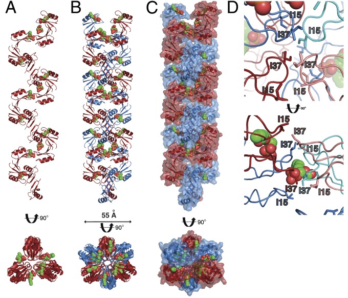
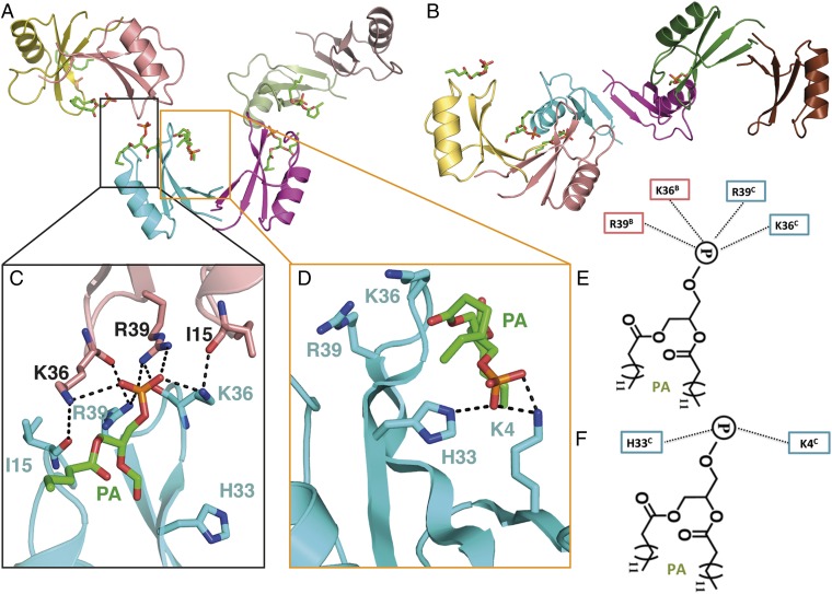
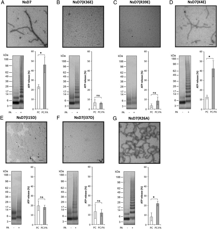
Defensins are cationic antimicrobial peptides that serve as important components of host innate immune defenses, often by targeting cell membranes of pathogens. Oligomerization of defensins has been linked to their antimicrobial activity; however, the molecular basis underpinning this process remains largely unclear. Here we show that the plant defensin NsD7 targets the phospholipid phosphatidic acid (PA) to form oligomeric complexes that permeabilize PA-containing membranes. The crystal structure of the NsD7-PA complex reveals a striking double helix of two right-handed coiled oligomeric defensin fibrils, the assembly of which is dependent upon the interaction with PA at the interface between NsD7 dimers. Using site-directed mutagenesis, we demonstrate that key residues in this PA-binding site are required for PA-mediated NsD7 oligomerization and coil formation, as well as permeabilization of PA-containing liposomes. These data suggest that multiple lipids can be targeted to induce oligomerization of defensins during membrane permeabilization and demonstrate the existence of a "phospholipid code" that identifies target membranes for defensin-mediated attack as part of a first line of defense across multiple species.

Keywords: antimicrobial peptides; defensins; host–pathogen interactions; innate defense; phospholipids.


Conflict of interest statement

M.D.H. is a former Vice President of Research in Hexima Ltd.

Figures

Fig. 1.
Fig. 1.
NsD7 binds PA and forms oligomers. (A) NsD7 forms oligomers in the presence of PA as determined by protein–protein cross-linking with BS3 followed by SDS/PAGE and Coomassie Brilliant Blue staining. (B) TEM of NsD7:PA complexes, NsD7 alone, or PA alone. (Scale bars, 100 nm.)
Fig. S1.
Fig. S1.
Sequence alignment of NsD7 with plant defensins NaD1 (AF509566, from ornamental tobacco), TPP3 (AAA80496, from tomato), and MtDef4 (2LR3, from barrel medic). The GenBank accession numbers and plant sources are given in parentheses. Residues that are identical to NsD7 are shaded in black, and similar residues are shaded in gray. The conserved cysteine residues involved in disulfide bonds are indicated by connecting lines, and the percent amino acid sequence identity relative to NsD7 is shown. The numbering above the sequences is relative to NsD7.
Fig. 2.
Fig. 2.
The NsD7:PA complex forms a double-stranded helix. (A) The NsD7:PA complex adopts a right-handed coiled structure. The coil shown comprises the content of three asymmetric units. PA molecules are shown as green (carbon), orange (phosphate), and red spheres (oxygen). (B and C) Two neighboring NsD7:PA coils form a defensin double helix. (D) The interior of the NsD7:PA oligomer harbors a series of intercalating isoleucine residues (I15 and I37). Top view is down the center of the oligomer; Bottom view is the same view rotated 90° on the horizontal axis.
Fig. 3.
Fig. 3.
Crystal structure of the NsD7:PA complex. (A) Three NsD7 dimers are arranged in a tip-to-tip configuration, with each neighboring dimer positioned orthogonally from its neighbor. A total of six PA molecules are bound by the three NsD7 dimers. (B) An identical view as in A, rotated 90° around the horizontal axis. The arrangement of the dimers leads to an S-shaped trimer of dimers. (C) Type I and (D) type II PA-binding sites. Hydrogen bonds or ionic interactions are marked as black dashed lines. Schematic representation of the (E) type I and (F) type II PA-binding sites.
Fig. 4.
Fig. 4.
Structure–function analysis of the interaction between NsD7 and PA. (A) Wild-type NsD7, type I PA-binding site mutants (B) NsD7(K36E) and (C) NsD7(R39E), type II PA-binding site mutant (D) NsD7(K4E), isoleucine zipper mutants (E) NsD7(I15D) and (F) NsD7(I37D), and interoligomer fibril formation mutant (G) NsD7(R26A). Each subpanel displays data for defensin oligomerization in the presence of PA as assessed by TEM (Top) or biochemical cross-linking using BS3 followed by SDS/PAGE and Coomassie Brilliant Blue staining (Bottom Left) and liposome permeabilization as measured by percentage of ATP release (relative to a Triton-X100 control) from liposomes comprising PC or PC:PA (Bottom Right). Graphs are representative of at least three independent experiments. Error bars, SEM. *P < 0.05; ns, not significant. (Scale bars, 100 nm.)
Fig. 5.
Fig. 5.
Oligomeric NsD7:PA and NaD1:PIP2 complexes have distinct assembly mechanisms. (A) Cartoon diagram of NsD7:PA complex illustrating the tip-to-tip dimer–dimer arrangement in the oligomer mediated by interaction with PA (dimer 1 in cyan and magenta and dimer 2 in sand and green). The key residues comprising the PA-binding site are shown as sticks; PA is shown as green sticks. (B) Cartoon diagram of NaD1:PIP2 complex illustrating the side-to-side dimer–dimer arrangement in the oligomer mediated by interaction with PIP2 (dimer 1 in cyan and magenta and dimer 2 in sand and green). The view of the cyan and magenta dimer 1 is conserved from A. Key residues comprising the PIP2 binding sites are shown as sticks, and PIP2 molecules are shown as sticks. (C) Schematic space-fill diagram of NsD7–PA oligomer assembly. NsD7 dimers are colored as in A with one dimer–dimer unit in the assembly highlighted by a red boundary; PA molecules are shown as black circles. (D) Schematic space-fill diagram of NaD1-PIP2 oligomer assembly. NaD1 dimers are colored as in B with one dimer–dimer unit in the-assembly highlighted by a red boundary; PIP2 molecules are shown as black circles.

References

    1. Brogden KA. Antimicrobial peptides: Pore formers or metabolic inhibitors in bacteria? Nat Rev Microbiol. 2005;3(3):238–250. - PubMed
    1. Chu H, et al. Human α-defensin 6 promotes mucosal innate immunity through self-assembled peptide nanonets. Science. 2012;337(6093):477–481. - PMC - PubMed
    1. Poon IKh, et al. Phosphoinositide-mediated oligomerization of a defensin induces cell lysis. eLife. 2014;3:e01808. - PMC - PubMed
    1. Athenstaedt K, Daum G. Phosphatidic acid, a key intermediate in lipid metabolism. Eur J Biochem. 1999;266(1):1–16. - PubMed
    1. Testerink C, Munnik T. Phosphatidic acid: A multifunctional stress signaling lipid in plants. Trends Plant Sci. 2005;10(8):368–375. - PubMed

Publication types

Associated data