. 2016 Oct 4;113(40):11283-11288.
doi: 10.1073/pnas.1607606113. Epub 2016 Sep 19.

Whole-exome sequencing identifies multiple loss-of-function mutations of NF-κB pathway regulators in nasopharyngeal carcinoma

Affiliations

Whole-exome sequencing identifies multiple loss-of-function mutations of NF-κB pathway regulators in nasopharyngeal carcinoma

Hong Zheng et al. Proc Natl Acad Sci U S A. .

Abstract

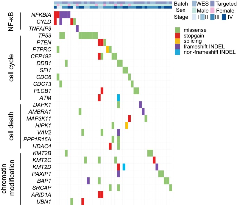
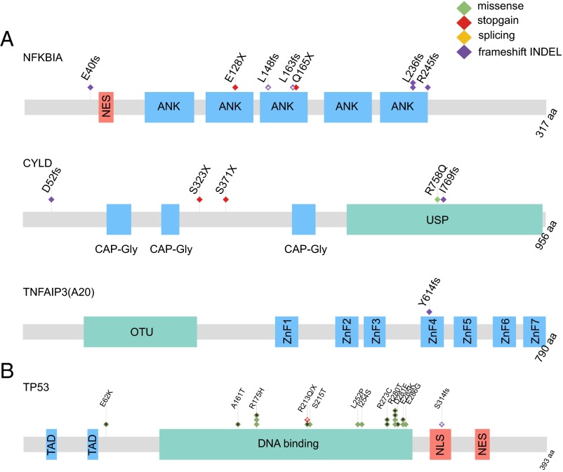
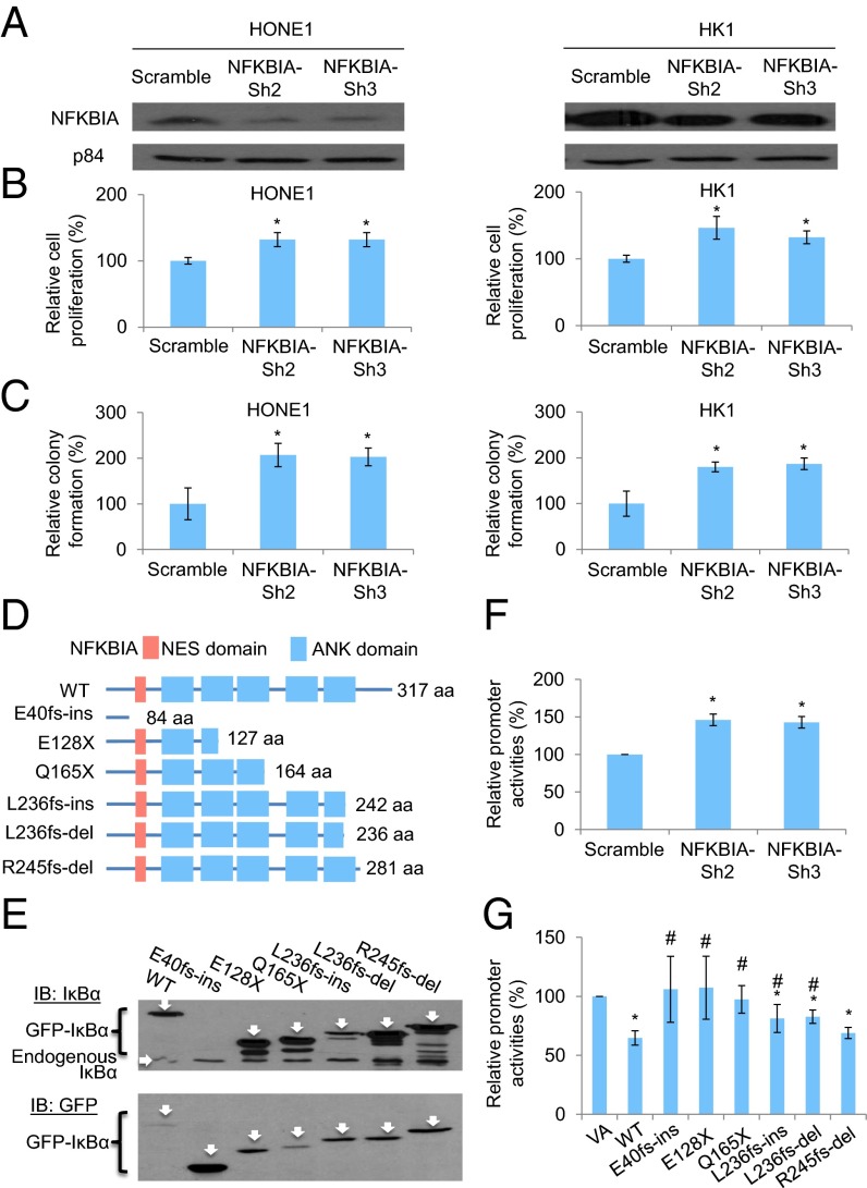
Nasopharyngeal carcinoma (NPC) is an epithelial malignancy with a unique geographical distribution. The genomic abnormalities leading to NPC pathogenesis remain unclear. In total, 135 NPC tumors were examined to characterize the mutational landscape using whole-exome sequencing and targeted resequencing. An APOBEC cytidine deaminase mutagenesis signature was revealed in the somatic mutations. Noticeably, multiple loss-of-function mutations were identified in several NF-κB signaling negative regulators NFKBIA, CYLD, and TNFAIP3 Functional studies confirmed that inhibition of NFKBIA had a significant impact on NF-κB activity and NPC cell growth. The identified loss-of-function mutations in NFKBIA leading to protein truncation contributed to the altered NF-κB activity, which is critical for NPC tumorigenesis. In addition, somatic mutations were found in several cancer-relevant pathways, including cell cycle-phase transition, cell death, EBV infection, and viral carcinogenesis. These data provide an enhanced road map for understanding the molecular basis underlying NPC.

Keywords: APOBEC-mediated signature; NF-κB signaling; nasopharyngeal carcinoma; somatic mutation landscape; whole-exome sequencing.


Conflict of interest statement

The authors declare no conflict of interest.

Figures

Fig. 1.
Fig. 1.
Somatic mutation rates and signatures in NPC. (A) Number of nonsilent somatic mutations in NPC tumors. (B) Trinucleotide contexts of somatic mutations occurring at cytosine nucleotides in NPC. The font sizes of the nucleotides at the 3′ and 5′ positions are proportional to their frequencies. (C) Mutational signatures are displayed according to 96 substitution classification defined by the substitution class and sequence context immediately before and after the mutated base (9). Vertical axis displays percentage of mutations attributed to a specific mutation type. The trinucleotide contexts of the mutated bases are shown on the x axis. (D) Expressions of APOBEC3A and APOBE3B evaluated by qRT-PCR in an additional 22 tumor pairs.
Fig. 2.
Fig. 2.
Landscape of somatic mutations in NPC. The matrix shows important mutated genes and their associated pathways in NPC primary tumors analyzed by WES and targeted resequencing. Genes are shown on left-hand side. Only genes with detectable expression in RNASeq analysis are shown. Each column represents one tumor.
Fig. 3.
Fig. 3.
Significantly mutated genes and relevant pathway in NPC. (A) Schematics of NF-κB pathway genes NFKBIA, CYLD, and TNFAIP3 showing the positions of individual somatic mutations identified in NPC. (B) Schematics of TP53 mutations. Each diamond represents one tumor. Protein domain and region information is obtained from the UniProt database. ANK, ankyrin repeat; CAP-Gly, cytoskeleton-associated proteins–Gly-rich domain; fs, frameshift mutation; NES, nuclear export signal; NLS, nuclear localization signal; OTU, ovarian tumor domain; TAD, transactivating domain; USP, ubiquitin-specific protease domain; X, stopgain mutation; ZnF1-7, Zinc finger domain 1–7. *Somatic mutations from previous WES studies (4, 12).
Fig. 4.
Fig. 4.
Functional investigation of NFKBIA knockdown in NPC cells and the truncating mutations identified in the IκBα protein. (A) Expression of IκBα protein in both NPC HONE1 and HK1 cell lines after knockdown with two independent sets of shRNA knockdown oligonucleotides. (B) Cell proliferation assay of HONE1 and HK1 cells after NFKBIA knockdown. The relative cell proliferation rate was compared with the corresponding scramble control. (C) Colony formation assay of HONE1 and HK1 cells after NFKBIA knockdown. (D) Schematic diagram illustrates the truncated site of each mutant of IκBα protein by in silico analysis. NES, nuclear export signal. (E) Western blot results illustrate the protein size of each mutant. The IκBα was tagged with an N-terminal GFP tag. The GFP-IκBα-WT (61.9 kDa), GFP-E40fs-ins (35.659 kDa; this mutant lost the epitope for the IκBα antibody recognition; therefore, it cannot be detected by the IκBα-specific antibody), GFP-E128X (41.68 kDa), GFP-Q165X (45.49 kDa), GFP-L236fs-ins (53.84 kDa), GFP-L236fs-del (53.04 kDa), and GFP-R245fs-del (57.85 kDa) were detected by specific antibody against the (Upper) IκBα and (Lower) GFP. IB, immunoblot. (F) NF-κB–specific dual luciferase promoter assay in the 293T cell lines with NFKBIA knockdown. (G) NF-κB–specific dual luciferase promoter assay in the 293T cell line with expression of IκBα-WT and mutants. This cell line has constitutive NF-κB activation. The relative promoter activity value was compared with the VA control. The data shown in B, C, F, and G represent means ± SD (n = 3). *Statistical significance (P < 0.05) compared with the scramble control or VA; #statistical significance (P < 0.05) compared with the WT.

References

    1. Bruce JP, Yip K, Bratman SV, Ito E, Liu FF. Nasopharyngeal cancer: Molecular landscape. J Clin Oncol. 2015;33(29):3346–3355. - PubMed
    1. Lo KW, Chung GT, To KF. Deciphering the molecular genetic basis of NPC through molecular, cytogenetic, and epigenetic approaches. Semin Cancer Biol. 2012;22(2):79–86. - PubMed
    1. Dai W, et al. Comparative methylome analysis in solid tumors reveals aberrant methylation at chromosome 6p in nasopharyngeal carcinoma. Cancer Med. 2015;4(7):1079–1090. - PMC - PubMed
    1. Lin DC, et al. The genomic landscape of nasopharyngeal carcinoma. Nat Genet. 2014;46(8):866–871. - PubMed
    1. Lawrence MS, et al. Mutational heterogeneity in cancer and the search for new cancer-associated genes. Nature. 2013;499(7457):214–218. - PMC - PubMed

Publication types