

Condition medium of HepG-2 cells induces the transdifferentiation of human umbilical cord mesenchymal stem cells into cancerous mesenchymal stem cells
- PMID: 27648133
- PMCID: PMC5009395
Condition medium of HepG-2 cells induces the transdifferentiation of human umbilical cord mesenchymal stem cells into cancerous mesenchymal stem cells
Abstract
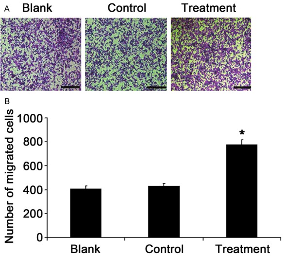
This study aimed to investigate the transdifferentiation of human umbilical cord mesenchymal stem cells (hUCMSCs) into cancer-associated mesenchymal stem cells (CA-MSCs) after incubation with condition medium (CM) from liver cancer HepG-2 cells, and the biobehaviors (proliferation and migration) of these CA-MSCs were further evaluated. The supernatant of HepG-2 cells was collected and mixed with equal volume of low glucose DMEM. The resultant medium was used to treat hUCMSCs for 48 h. The expression of CA-MSCs related proteins and miR-221 was detected in cells. The supernatant of induced hUCMSCs was mixed with equal volume of high glucose DMEM, and the resultant medium was used treat HepG-2 cells for 48 h and the proliferation and migration of HepG-2 cells were evaluated. Moreover, HepG-2 cells were co-cultured with hUCMSCs and then the proliferation and migration of HepG-2 cells were assessed. After incubation with the supernatant from HepG-2 cells, hUCMSCs showed significantly elevated expression of vimentin, fibroblast activation protein (FAP) and miR-221. The supernatant of induced hUCMSCs was able to significantly increase the proliferation and migration of HepG-2 cells. Following co-culture, the proliferation and migration of HepG-2 cells increased dramatically. These findings suggest that the supernatant of HepG-2 cells is able to induce the phenotype of CA-MSCs and the supernatant of CA-MSCs may promote the proliferation and migration of HepG-2 cells. These findings provide experimental evidence for the cellular remodeling in tumor microenvironment and the safety of clinical use of hUCMSCs.
Keywords: Stem cells; cell migration; hepatocellular carcinoma; transdifferentiation; umbilical cord mesenchymal stem cells.
Figures


References
-
- Moon HS. Chemopreventive Effects of Alpha Lipoic Acid on Obesity-Related Cancers. Ann Nutr Metab. 2016;68:137–144. - PubMed
-
- Albini A, Sporn MB. The tumour microenvironment as a target for chemoprevention. Nat Rev Cancer. 2007;7:139–147. - PubMed
-
- Studeny M, Marini FC, Champlin RE, Zompetta C, Fidler IJ, Andreeff M. Bone marrow-derived mesenchymal stem cells as vehicles for interferon-beta delivery into tumors. Cancer Res. 2002;62:3603–3608. - PubMed
-
- Studeny M, Marini FC, Dembinski JL, Zompetta C, Cabreira-Hansen M, Bekele BN, Champlin RE, Andreeff M. Mesenchymal stem cells: potential precursors for tumor stroma and targeted-delivery vehicles for anticancer agents. J Natl Cancer Inst. 2004;96:1593–1603. - PubMed
LinkOut - more resources
Full Text Sources
Miscellaneous